
Official websites use .gov
A .gov website belongs to an official government organization in the United States.
Secure .gov websites use HTTPS
A lock (
) or https:// means you’ve safely connected to the .gov website. Share sensitive information only on official, secure websites.
Software Tools
The Biosystems and Biomaterials Division (BBD) develops measurement technologies to bolster the bioeconomy and foster innovation. To support these efforts, BBD staff have developed open-source software tools to address critical vacancies in the available analysis landscape across a broad portfolio of activities. Note that the software products listed below are not considered Standard Reference products and provided as is. Please see the NIST Software Disclaimer statement for additional information.
IMAGE ANALYSIS
| Image | Name | Description | Contact |
|---|---|---|---|

| DiameterJ (available but no longer supported) | DiameterJ is a validated nanofiber diameter characterization tool for use in ImageJ [1/2] and Fiji. DiameterJ is able to analyze an image and find the diameter of nanofibers or microfibers at every pixel along a fibers axis and produces a histogram of these diameters. Included with this histogram are summary statistics such as mean fiber diameter and most occurring fiber diameter. | Lin |

| SQuIRE, CARPE (available but no longer supported) | CARPE is an ImageJ plugin to convert bright-field microscope images into absorbance images. SQuIRE is a micromanager plugin for automated acquisition of the images. | Lin |

| CRIKit2 | CRIKit2 is a Python library for processing hyperspectral imagery, primarily from coherent anti-Stokes Raman scattering (CARS) microscopy systems. This library contains command line tools, interactive widgets, and a user interface. | Camp |
| NIST developed a procedure for characterizing the performance of a fluorescence microscope by benchmarking the detection threshold, saturation, and linear dynamic range to a physical artifact, such as a fluorescent material. This method has already been implemented by several universities and companies, and is proven to improve the reproducibility of wide-field fluorescence imaging. Our strategy adds value to image data repositories by benchmarking the conditions under which the images were acquired. | Asmar | |

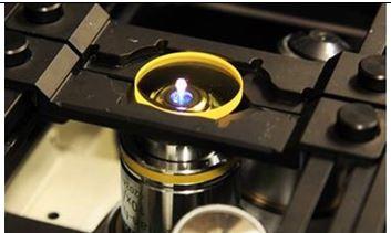
| WIPP | Several advanced microscopes have the capability to acquire overlapping image tiles automatically. Such automated acquisitions provide a way to image a large spatial coverage of a specimen, to take measurements at multiple physical length scales, and to learn the underlying models that describe the specimen. There is a need to assist imaging scientists with computational solutions that convert raw image tiles to calibrated, stitched, and viewable Giga-pixel images with interactive and traceable measurements and modeling tools. In other words, a need for a solution to go From Image Tiles to Web-Based Traceable Measurements and Interactive Modeling in One Stop (denoted as web image pipeline for interactive discoveries). | Asmar |
Credit:
NIST
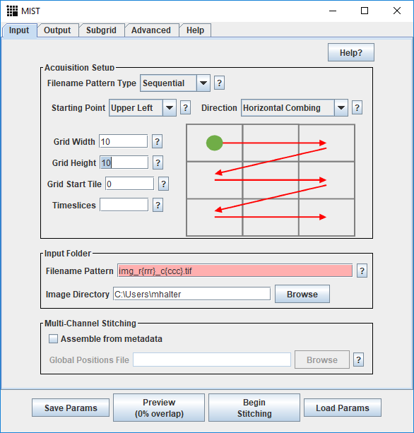
| MIST | Microscopy Image Stitching Tool (MIST), is a stitching tool for 2D grids of images. MIST estimates the stage mechanical model (actuator backlash, stage repeatability ‘r’, etc.) from computed pairwise translations and then minimizes stitching errors by optimizing the translations within a (4r)^2 square area. This minimizes the maximum uncertainty related to the translation computation for any pair of images. | Asmar |

| EGT | EGT is a novel and empirically derived image gradient threshold selection method for separating foreground and background pixels in an image. The method works across multiple optical microscopy imaging modalities and cell lines. | Asmar |

| Fogbank | FogBank separates cells when they are confluent and touching each other. The separation of touching cells in microscopy images is critical for the counting, identification and measurement of individual cells. | Asmar |
Genomic analysis
| Image | Name | Description | Contact |
|---|---|---|---|

| GIAB Integration Pipeline | Genome In A Bottle small and structural variant integration pipeline for generating GIAB benchmark sets. | Olson |
| metagenomeFeatures | R packages for working with 16S rRNA databases. | Olson |
MACHINE LEARNING
| Image | Name | Description | Contact |
|---|---|---|---|

| pyMCR is a small Python 3 package for performing multivariate curve resolution. Currently, it implements a simple alternating regression scheme (MCR-AR). The most common implementation is with ordinary least-squares regression, MCR-ALS. |
Engineering Biology
| Image | Name | Description | Contact |
|---|---|---|---|
| LANTERN is an intrinsically interpretable machine-learning approach that enables quantitative prediction of the effects of multiple mutations to a protein’s function. LANTERN’s predictive accuracy equals or exceeds the accuracy from alternative approaches, including deep neural networks. At the same time, LANTERN extracts useful insights into the global structure and biophysics of a protein’s sequence-function relationships. | |||
| ctRSD-simulator-2.0 was designed to be a comprehensive, scalable, and user-friendly model for simulating the kinetics of cotranscriptionally encoded RNA strand displacement (ctRSD) circuits. The goal of the simulator is to aid the user to design, predict, and understand the behavior of ctRSD circuits. Also, providing experimentalists with a functional model allowing for the efficient in silico prototyping of ctRSD systems. In addition to the kinetic simulations, the simulator package also has a sequence compiler function that can be used to assemble the DNA sequences necessary to test a specific component / design in experiments. |
DISCLAIMER
For more information concerning software licenses and use, please see the NIST Software Disclaimer statement.