
Official websites use .gov
A .gov website belongs to an official government organization in the United States.
Secure .gov websites use HTTPS
A lock (
) or https:// means you’ve safely connected to the .gov website. Share sensitive information only on official, secure websites.
3D Cell-Scaffold Interactions
Summary
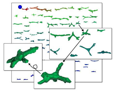
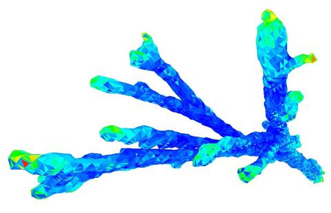
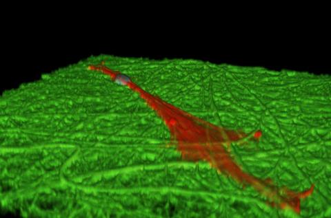
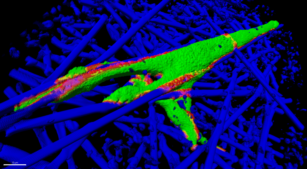
When adherent cells are cultured in tissue culture plates, they adhere to a planar surface. In native tissue in vivo, cells often exist within a three-dimensional extracellular matrix that surrounds them on all sides. 3D scaffolds are being used to provide a more realistic in vitro culture environment and as 3D templates for tissue regeneration. The chemical, structural, and mechanical properties of the scaffold influence cell function, and these effects are at least partially dependent on the morphologies that cells take on during scaffold culture. We are imaging cells in different types of scaffolds in order to assess how scaffold culture influences the 3D shape of cells. We collect 3D image data from large numbers of cells by confocal microscopy and use computational methods to process, segment, and analyze 3D cell shape. This work may provide an understanding of how scaffold properties can be used to direct function through modulation of cell morphology.
Publications
- Florczyk SJ, Hotaling NA, Simon M, Chalfoun J, Horenberg AL, Schaub NJ, Wang D, Szczypiński PM, DeFelice VL, Bajcsy P, Simon Jr CG (2023) Measuring Dimensionality of Cell-Scaffold Contacts of Primary Human Bone Marrow Stromal Cells Cultured on Electrospun Fiber Scaffolds. Journal of Biomedical Materials Research Part A 111(1), 106-117. https://doi.org/10.1002/jbm.a.37449
- Pazmino Betancourt BA, Florczyk SJ, Simon M, Juba D, Douglas JF, Keyrouz W, Bajcsy P, Lee C, Simon Jr CG (2018) Effect of the scaffold microenvironment on cell polarizability and capacitance determined by probabilistic computations. Biomedical Materials 13, 025012.
- Florczyk SJ, Simon M, Juba D, Pine PS, Sarkar S, Chen D, Baker PJ, Bodhak S, Cardone A, Brady MC, Bajcsy P, Simon Jr CG (2017) A bioinformatics 3D cellular morphotyping strategy for assessing biomaterial scaffold niches. ACS Biomaterials Science & Engineering 3, 2301-2313.
- Tutak W, Jyotsnendu G, Bajcsy P, Simon Jr CG (2017) Measuring the 3D shapes of organelles in stem cells cultured on nanofiber scaffolds. Journal of Biomedical Materials Research: Part B - Applied Biomaterials 105, 989-1001..
- Chen D, Sarkar S, Candia J, Florczyk SJ, Bodhak S, Driscoll MK, Simon Jr CG, Dunkers JP, Losert W (2016) Machine learning based methodology to identify cell shape phenotypes associated with microenvironmental cues. Biomaterials 104, 104-118.
- Bajcsy P, Cardone A, Chalfoun J, Halter M, Juba D, Kociolek M, Majurski M, Peskin A, Simon Jr CG, Simon M, Vandecreme A, Brady M (2015) Survey statistics of automated segmentations applied to optical imaging of mammalian cells. BMC Bioinformatics 16, 330, 1-28.
- Bajcsy P, Simon M, Florczyk S, Simon Jr CG, Juba D, Brady M. (2015) A method for the evaluation of thousands of automated 3D stem cell segmentations. Journal of Microscopy 260, 363-376.
- Sarkar S, Baker BA, Chen D, Pine PS, McDaniel JH, Salit ML, Losert W, Simon Jr CG, Dunkers J (2016) Roles of nanofiber scaffold structure and chemistry in directing human bone marrow stromal cell response. Advances in Tissue Engineering & Regenerative Medicine 1, 00003.
- Farooque TM, Camp Jr CH, Tison CK, Kumar G, Parekh SH, Simon Jr CG (2014) Measuring stem cell dimensionality in tissue scaffolds. Biomaterials 35, 2558-2567.
- Juba D, Cardone A, Ip CY, Simon Jr CG, Tison CK, Kumar G, Brady M, Varshney A (2013) Parallel geometric classification of stem cells by their three dimensional morphology. Computational Science & Discovery 6, 015007.
- Kumar G, Tison CK, Chatterjee K, Pine PS, McDaniel JH, Salit ML, Young MF, Simon Jr CG (2011) The determination of stem cell fate by 3D scaffold structures through the control of cell shape. Biomaterials 32, 9188-9196.
- Dorsey SM, Lin-Gibson S, Simon Jr CG (2009) X-ray microcomputed tomography for the measurement of cell adhesion and proliferation in polymer scaffolds. Biomaterials 30, 2967–2974.
Contributors
Carl Simon, Stephanie Florczyk, Peter Bajcsy, Nathan Hotaling, Nicholas Schaub, Mylene Simon, Jack Douglas, Beatriz Pazmino