Official websites use .gov
A .gov website belongs to an official government organization in the United States.
Secure .gov websites use HTTPS
A lock (
) or https:// means you’ve safely connected to the .gov website. Share sensitive information only on official, secure websites.
Standards for Nuclear Medicine
Tens of millions of U.S. citizens annually undergo medical imaging scans that use radioactive tracers to highlight areas of interest in the body. The most frequent diagnostic procedures are scans of the heart, lungs and bone using short-lived radioactive atoms such as technetium-99m (six-hour half-life). A newer set of procedures for brain imaging uses sugars labeled with radioactive atoms such as fluorine-18 (110-minute half-life) that release particles called positrons (positively charged electrons). These positron emission tomography (PET) scans show sugar uptake in different regions of the brain, which is valuable for monitoring the conditions of stroke victims.
In the 1970s, chemists and physicists at the National Bureau of Standards (NBS, now the National Institute of Standards and Technology, or NIST) worked with the Food and Drug Administration (FDA) and U.S. pharmaceutical manufacturers to establish a measurement assurance program for nuclear medicine. The program ensured that all the radioactive isotopes in commercial use in the U.S. linked back to radioactivity standards developed in Building 245. Today, industry research associates working alongside NIST staff produce several radiopharmaceutical tracer Standard Reference Materials (SRMs) per year that are used by U.S. companies in their quality assurance programs.
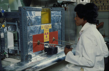
Using short-lived radioisotopes in imaging also requires a rapid and accurate method of ensuring that each dose will be the same. NIST pioneered the modern methods of standardizing these short-lived radionuclides, and NIST and Centro Español de Metrologia (CEM), Spain’s measurement science institute, developed a technique known as the CIEMAT-NIST method that is used to calibrate many medical radionuclides.
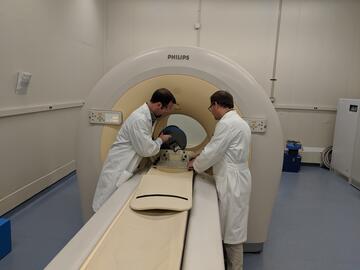
The rapid expansion of PET scanning facilities in the U.S. and the ability to map PET scans onto anatomical three-dimensional magnetic resonance imaging (MRI) or X-ray computed tomography (CT) scans has led to the need for phantoms, materials that simulate human tissues, that can be used to calibrate PET scanners in three dimensions as well. The addition of a PET/CT system in 2010 allowed NIST to begin developing phantoms and other imaging standards for positron-emitting radiopharmaceuticals, which has had an international impact.