

Official websites use .gov
A .gov website belongs to an official government organization in the United States.

Secure .gov websites use HTTPS
A lock (
) or https:// means you’ve safely connected to the .gov website. Share sensitive information only on official, secure websites.


Label-Free Light Scattering Microscopy for Gene Therapy Quality Control
Summary
Gene therapies hold the potential to treat previously incurable conditions but manufacturing complex viral vectors and nanoparticles reliably and affordably remains a critical hurdle. NIST is developing single-particle measurement technologies to help manufacturers evaluate particle quality, structure, and consistency directly during production.
Description
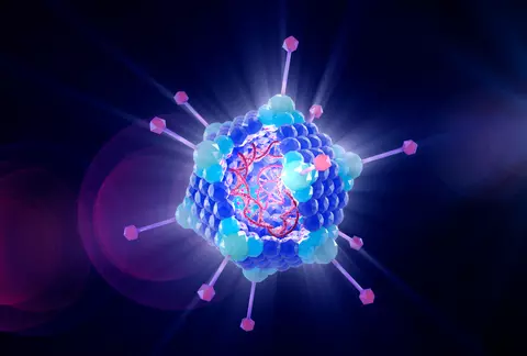
What does this gene delivery particle contain?
Why This Matters Gene therapies rely on nanometer-scale delivery vehicles, such as adenoviral and lentiviral vectors, to transport corrective genetic material into patient cells. While early treatments have achieved breakthroughs in treating rare inherited conditions, expanding these therapies into broader diseases (such as cancer and muscular disorders) requires rapid, scalable biomanufacturing processes.
During biomanufacturing of viral vectors, batches often contain a mixture of functional particles, empty shells, partially filled vectors, and unwanted aggregates. Current analytical methods used to assess these populations are often slow, expensive, consume large sample volumes, and cannot provide real-time process control. This measurement bottleneck contributes to high treatment costs and extended development timelines.
To address these metrology needs, NIST is developing continuous, multi-attribute light scattering capabilities to rapidly measure individual particle mass, size, concentration, and composition without destructive sampling or chemical labels.
How It Works When laser light interacts with an individual nanoparticle, the scattered light pattern encodes details about the particle's physical properties, size, and internal mass. By capturing and analyzing this scattered light on a particle-by-particle basis using high-resolution optics, thousands of individual particles can be measured rapidly in their native state.
To bring this capability into real-world manufacturing workflows, our team is integrating light scattering microscopy with continuous microfluidic sampling and event-based sensing. This combined approach dramatically reduces data storage requirements while maintaining high throughput from active fluidic streams.
– Visit Gene Delivery Systems for broader program goals
Project Areas:
What We Are Working On: We are a multidisciplinary, collaborative team of researchers with expertise across five critical project areas
Contact us at gtp [at] nist.gov (Gene Therapy Particles)
Optics and Microscopy Instrumentation
Our current research efforts include:
- Designing and constructing custom laser-based microscope systems for quantitative light-scatter measurements of individual bio-nanoparticles.
- Employing advanced image analysis techniques to accurately measure particle loading, size, and concentration.
- For further information about this project, contact Alexander Peterson or Aaron Goldfain
- NRC positions here:
- PREP position here:
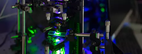
Optical Modeling
Our team's research efforts include:

- Applying sophisticated optical models to interpret and understand image-based particle signals.
- Developing computational models that extract physical properties from imaging data, laying the foundation for a comprehensive set of quality attributes.
- Advancing light-scatter nanoscopy techniques to characterize gene delivery systems and complex bio-nanoparticles.
- NRC position here: Light-Scatter Nanoscopy of Gene Delivery Systems and Complex Bio-Nanoparticles
- For further information about this project, contact Aaron Goldfain or Thomas Germer
Viral Vectors
Our team's current research efforts include:

- Developing upstream and downstream processes for the production and purification of viral vectors in bioreactors.
- Utilizing conventional and state-of-the-art methods to analyze viral vector titer and purity throughout the manufacturing process.
- Designing orthogonal measurement assays to compare novel light scattering techniques with current industry standards.
- Integrating microfluidic and optical measurements with on-line bioreactor monitoring, in collaboration with the Gene Delivery Systems program.
- To learn more about this project, contact Edward Kwee
Computational Image Analysis

- Use neuromorphic sensing (event cameras) for faster temporal sampling and higher data transfer from the sensor to a computer
- Research event data representations and quality metrics suitable for low-light microscope imaging of the stochastic motion of nanoparticles
- Develop computational workflows to derive hydrodynamic diameters from event data and evaluate their accuracies
- Explore neuromorphic nonlinear models for centroid localization and tracking of nanoparticles
- Research enablers of high-throughput high-confidence measurements in bioreactors using novel cameras and computational hardware
- Contact Peter Bajcsy for more information.
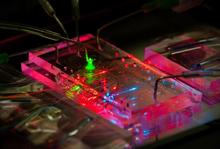
Microfluidic Devices
Our team is actively working to:
- Design, create, and operate microfluidic devices that precisely control the flow and filtration of nanoscale viral particles.
- Ensure the reliability and accuracy of particle positioning over imaging sensors through advanced microfluidic techniques.
- Develop chip-based sensing systems for in-line and on-line particle measurements..
- To learn more about this project, contact Greg Cooksey or Matthew DiSalvo
- Learn about our microfluidic and cytometry projects with the NRC postdoctoral fellowship program