
Official websites use .gov
A .gov website belongs to an official government organization in the United States.
Secure .gov websites use HTTPS
A lock (
) or https:// means you’ve safely connected to the .gov website. Share sensitive information only on official, secure websites.
Summary
Super-resolution optical microscopy extends deep into the nanometer scale with high throughput. The Nanostructure Fabrication and Measurement Group is improving accuracy far beyond the diffraction limit and applying the resulting measurement capabilities to diverse applications including photonic fabrication, microsystem characterization, and nanoparticle metrology.
Description
Optical microscopy has been fundamental to science for four centuries, enabling resolution of the microscale. In the last two decades, scientists have made the remarkable discovery that super-resolution methods can extend optical microscopy deep into the nanoscale. It is now possible to detect and localize objects as small as a single molecule with subnanometer precision, in comparison to the conventional resolution limit of a few hundred nanometers. Analysis of optical intensity variation through focus is then a rich source of information about nanostructure positions, orientations, and dimensions. Driven by these new opportunities, the microscopy community has focused their attention on developing methods, improving precision, and pursuing applications. However, this has left the development of accuracy and traceability as a critical problem for NIST to solve. Reliable data are necessary to make quantitative comparisons between studies and to put ordinary instruments to best use by achieving accurate and efficient measurements throughout wide and deep focal volumes. Moreover, advances in imaging in three dimensions often present additional sources of error, further motivating better accuracy.
The root cause of the problem is that standards and calibrations to achieve accuracy at the relevant scales are lacking. To address this issue, we are applying nanofabrication to develop microscopy standards and calibration methods that dramatically improve the accuracy of optical microscopy beyond the diffraction limit. We then apply these methods to measure nanostructures in a virtuous cycle to improve fabrication processes, enabling novel devices and microscopy measurements.
We have advanced and applied super-resolution optical microscopy in diverse contexts, including: development of standards to identify and correct errors in optical microscopy; localization of single molecules to probe mechanical failure of composite materials; localization of single quantum dots for fabricating nanophotonic devices with high efficiency; localization of nanoparticles within nanostructured devices for colloidal metrology; tracking of single particles for measuring the effects of microfabrication tolerances on the motion complex microsystems at the nanoscale; through-focus optical microscopy for dimensional metrology of nanostructures and quality control of fabrication processes; and shadowgraphy for volumetric metrology of flying microdroplets for inkjet fabrication and aerosol epidemiology. Current research in the Division aims to fabricate standards for localization microscopy in three dimensions, establish traceability of standards and calibrations, and apply optical microscopy for characterization of nanofabrication processes, nanoparticle medicines, and nanoplastic pollutants.
Highlights
Highlight: NIST puts the optical microscope under the microscope
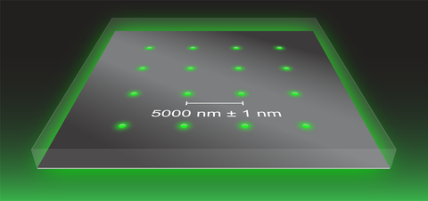
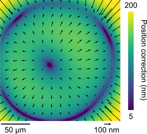
We are enabling the application of optical microscopy to localize single emitters, such as fluorescent molecules or particles, with accuracy approaching the atomic scale across an ultrawide field. Optical microscopes typically lack the calibration that is necessary to obtain accurate data at this scale. Localization microscopy can be precise, consistently indicating the same position of a single emitter to within one nanometer. However, this localization result can be inaccurate – the true location of the emitter can be up to several micrometers away due to systematic effects, primarily pertaining to magnification and distortion calibration, resulting in a discrepancy between precision and accuracy of up to four orders of magnitude. To solve this egregious but common problem, we developed a new method to correct these errors. Our method uses arrays of nanoscale apertures to provide reference positions for a Zernike field correction of aberration effects. Having calibrated our optical microscope using the aperture arrays, we reversed the process, using our microscope to identify imperfections in the prototype arrays from nanofabrication by electron-beam lithography and focused-ion-beam milling. The standards have the potential for mass production, and the ease and speed of optical microscopy enables rapid characterization of prototype standards and could facilitate quality control of standards for production and distribution to stakeholders.
- Copeland, C. R., Geist, J., McGray, C. D., Aksyuk, V. A., Liddle, J. A., Ilic, B. R., and Stavis, S. M., Subnanometer localization accuracy in widefield optical microscopy, Light: Science & Applications 7 (2018)
Highlight: Optical microscopy free from flatland
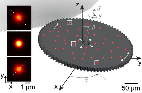
Conventional optical microscopes provide information about microscale samples in two dimensions – the lateral plane of a microscope slide. However, the third dimension – the axis perpendicular to the lateral plane – is just as important. We have developed a new method to convert the problematic effects of intrinsic aberrations, which affect most microscopes and arise from lens imperfections, into an innovative solution that enables conventional microscopes to accurately localize single emitters in all three dimensions. Our method relies on comprehensive calibration of how the shapes of emitter images change with position in the third dimension. Critically, the apparent positions of emitters in the first two dimensions also change with position in the third dimension, making localization in all three dimensions a prerequisite for accuracy. Our method accounts for these dependences, as well as how these dependences change with lateral position across an ultrawide imaging field. We tested our calibration method by tracking a constellation of fluorescent particles on the surface of a complex microsystem. Not only did this test confirm that the method is accurate in all three dimensions, it revealed complex motion of the gear in all six degrees of freedom, due to the effects of microfabrication tolerances on coupling interactions between parts of the system.
- Copeland, C. R., McGray, C. D., Ilic, B. R., Geist, J., and Stavis, S. M., Accurate Localization Microscopy by Intrinsic Aberration Calibration, Nature Communications 12 (2021)
Highlight: Avoiding the crack of doom
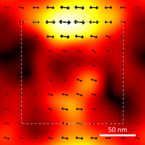
Just as a long journey begins with a single step, the deformations and fractures that cause catastrophic failure in materials begin with a few molecules out of place. This leads to a cascade of damage at larger scales, culminating in a total breakdown. That process is of urgent interest to researchers studying how to strengthen composite materials for critical components ranging from airplane wings and wind turbines to prosthetic joints. For many materials, it is difficult to see the early onset of damage because there are no visible markers to track its effects. We have developed a method to observe the effects of strain at the level of single molecules by measuring how a force changes the alignment of molecules in a polymer matrix. The technique uses super-resolution optical microscopy to measure the position and orientation of fluorescent molecules. We tested our method on a thin film of a polymer found in Lucite and Plexiglas that we doped with fluorescent molecules and deformed with nanoimprint lithography. Initially, the polymer was unstressed, and the fluorescent molecules were in random orientations. When we nanopatterned the polymer, deforming it in a specific direction, the fluorescent molecules followed the deformation, lining up with the path of the damage. Our study forms a basis for new applications of super-resolution microscopy for materials research.
- Wang, M., Marr, J. M., Davanco, M., Gilman, J. W., and Liddle, J. A., Nanoscale deformation in polymers revealed by single-molecule super-resolution location – orientation microscopy, Materials Horizons 6 (2019)