
Official websites use .gov
A .gov website belongs to an official government organization in the United States.
Secure .gov websites use HTTPS
A lock (
) or https:// means you’ve safely connected to the .gov website. Share sensitive information only on official, secure websites.
Description
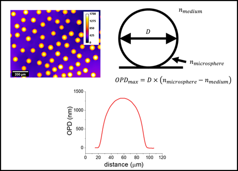
Using laboratory prepared microsphere samples to benchmark quantitative phase imaging systems.
Light microscopy is unparalleled as a quantitative technique for studying cell biology, but assuring that the results of measurements and analyses are accurate, repeatable and sharable is challenging. Here we provide information on reference materials, protocols and approaches for evaluating the reliability of imaging data and image analysis algorithms.
Reference materials and Protocols
| Image | Name | Description |
|---|---|---|

| Fluorescence Microscope Benchmarking | We have made available a method to characterize a fluorescence microscope's performance by benchmarking the detection threshold, saturation and linear dynamic range to a physical artifact (i.e. a fluorescent material). |

| ASTM F3294 - 18 Standard Guide for Performing Quantitative Fluorescence Intensity Measurements in Cell-based Assays with Widefield Epifluorescence Microscopy | Guidance document and checklist for performing relative intensity measurements for optical microscopy. |
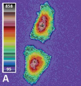
| Practical application of microsphere samples for benchmarking a quantitative phase imaging system | December 19, 2020: Author(s) Edward J. Kwee, Alexander W. Peterson, Michael Halter, John Elliott; Quantitative phase imaging (QPI) provides an approach for monitoring, for example, the growth rate of individual cells by measuring the optical pathlength of visible light. |
| Large Field of View Quantitative Phase Imaging of Induced Pluripotent Stem Cells and Optical Pathlength Reference Materials | February 23, 2018: Author(s) Edward J. Kwee, Alexander W. Peterson, Jeffrey R. Stinson, Michael W. Halter, Liya Yu, Michael P. Majurski, Joe Chalfoun, Peter Bajcsy, John T. Elliott; Induced pluripotent stem cells (iPSCs) are reprogrammed cells that can have heterogeneous biological potential. Quantitative imaging can help provide quality assurance metrics of reprogrammed iPSCs. |
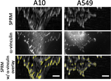
| Mass Measurements of Focal Adhesions in Single Cells Using High Resolution Surface Plasmon Resonance Microscopy | February 23, 2018: Authors(s) Alexander W. Peterson, Alessandro Tona, Michael W. Halter, Anne L. Plant, John T. Elliott; Surface plasmon resonance microscopy (SPRM) is a powerful label-free imaging technique with spatial resolution approaching the optical diffraction limit, and high sensitivity to small changes in index of refraction |
Image quality and Analytical assurance
| Image | Name | Description |
|---|---|---|
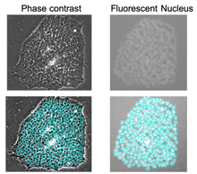
| Analyzing U-Net Robustness for Single Cell Nucleus Segmentation From Phase Contrast Images | July 28, 2020: Authors Chenyi Ling, Michael Majurski, Michael Halter, Jeffrey Stinson, Anne Plant, Joe Chalfoun; We quantify the robustness of the semantic segmentation model U-Net, applied to single cell nuclei detection, with respect to the following factors: (1) automated vs manual training annotations, (2) quantity of training data, and (3) microscope image focus. |
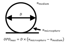
| Practical application of microsphere samples for benchmarking a quantitative phase imaging system | December 19, 2020: Author(s) Edward J. Kwee, Alexander W. Peterson, Michael Halter, John Elliott; Quantitative phase imaging (QPI) provides an approach for monitoring, for example, the growth rate of individual cells by measuring the optical pathlength of visible light. |
| Comparison of segmentation algorithms for fluorescence microscopy images of cells | June 14, 2011: Author(s) Alden A. Dima, John T. Elliott, James J. Filliben, Michael W. Halter, Adele P. Peskin, Javier Bernal, Marcin Kociolek, Mary C. Brady, Hai C. Tang, Anne L. Plant; Segmentation results from nine different segmentation techniques applied to two different cell lines and five different sets of imaging conditions were compared. Significant variability in the results of segmentation was observed. |
Image metadata efforts
- Open Microcopy Environment (OME) Bio-Formats is a software tool for reading and writing image data using standardized, open formats.
- 4d Nucleome MicroMeta provides a user interface to collect metadata about microscopes
External Organizations advancing measurement assurance for cell imaging
- Quality Assessment and Reproducibility for Instruments & Images in Light Microscopy (QUAREP-LiMi)
- ISAC / Cyto University Image Cytometer Characterization and Calibration, Lesson 1: Overview and Checklist for Benchmarking