Official websites use .gov
A .gov website belongs to an official government organization in the United States.
Secure .gov websites use HTTPS
A lock (
) or https:// means you’ve safely connected to the .gov website. Share sensitive information only on official, secure websites.
Traditionally, cell biology has been an academic subject area focused on the basic science of mammalian cells and single cell organisms like bacteria. Today, the quantitative application of cell biology is a segue to advanced manufacturing.
In the past couple of decades, the field of cell biology has incorporated many technological advances. These include whole genome sequencing, single cell RNA transcriptomics, sub-diffraction resolution of intracellular fluorescent probes, precision gene editing, and others. The “omics” methods and advances in large-scale cell imaging have enabled the accumulation of big datasets suitable for advanced mathematical analysis.
With these new tools, we are entering an era of cell biology where it will be possible to understand and predict the complex regulatory mechanisms that determine cell function, the “missing link” between basic science and useful therapeutics[i] and other bioeconomy products. With that understanding of the relationships between structure and function will come an increased ability to design, control, and manufacture cells.
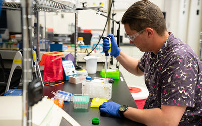
At NIST, we are developing systems that will lead to better quantification and prediction of complex biological processes for improved drug development, personalized medicine, cell therapies, and the design and manufacturing of cells and cell products for the health, agriculture, and environment sectors.
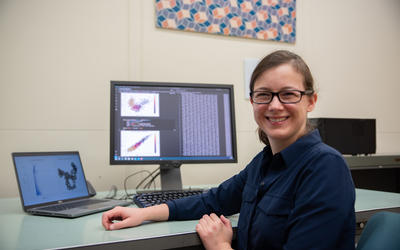
Selected Programs and Accomplishments
Quantifying dynamic biological processes with live cell imaging. Cells can respond to a combination of many extra-cellular signals, including chemical, mechanical, electrical and optical, and process these signals through multistep biochemical reactions. The stochastic nature of signal processing in cells results in a range of responses. Accurate characterization of a heterogeneous population of cells requires sampling a statistically significant number of individual cells. Ultimately, understanding the response functions of cells will allow us to predict the efficacy of a product or the outcome of a manufacturing process change. At NIST, high speed image data acquisition from living cells is one technology being used to collect the quantitative dynamic data needed to develop predictive models.
Thermodynamic principles describe and predict cell populations. Biological processes can be described by physical relationships, although much work is still needed in this area. We model the heterogeneity of cells as a population of microstates and apply thermodynamic principles for predicting population response.
Non-invasive stem cell imaging made possible with deep learning and very large image datasets. Imaging of living cells is a powerful tool for characterization and prediction since it provides quantitative data on morphological and chemical features of cells, as well as temporal and spatial information. Fluorescent labels and gene reporters provide a window into specific protein machinery of the cell but require radiation that can be damaging. We use fluorescent probes in cells to train neural networks to segment and track stem cells in culture that are imaged under nonfluorescent (bright field) illumination. This approach, in principle, will enable quality control of live, unlabeled cells with minimum perturbation during manufacturing.
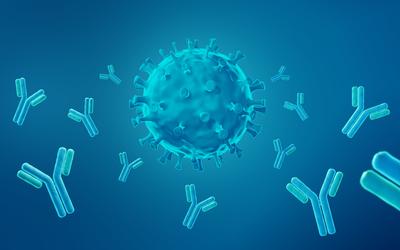
Improving Metrics for Cell-Based Therapies and Regenerative Medicines. This is one of the most exciting and challenging applications of advanced cell biology. Some cell-based therapies have been shown to cure, not just to treat, previously deadly cancers. At NIST, we are developing technologies that will enable better characterization of cell therapy products and processes. One approach uses the creation of reporter cell lines which are engineered to produce fluorescent reporter molecules when important control pathways are induced in cells. This approach will allow real time monitoring of a manufacturing protocol.
[i] Berger and Iyengar, 2009
News and Updates
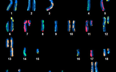
NIST Releases Trove of Genetic Data to Spur Cancer Research
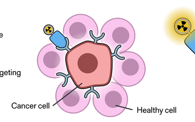
New NIST Standard Helps Deliver the Right Dosage of Cancer-Fighting Drugs
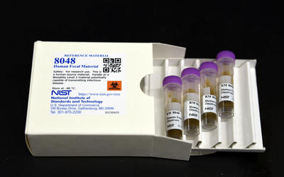
NIST Releases Reference Material to Aid Gut Microbiome Research
Blog Posts
Measure Twice, Cut Once: How Measuring Molecular ‘Scissors’ Can Further Groundbreaking Research
You Don’t Learn This in School: My Experience as a NIST Summer Intern