
Official websites use .gov
A .gov website belongs to an official government organization in the United States.
Secure .gov websites use HTTPS
A lock (
) or https:// means you’ve safely connected to the .gov website. Share sensitive information only on official, secure websites.
X-ray Methods for Chemical Imaging at Micro to Mesoscales
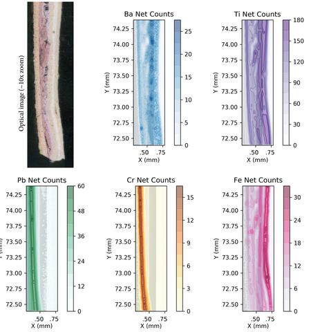
Lead content in residential wall paint: 10x light microscope[Left] with corresponding Micro XRF composition maps showing ~20 micrometer features.
Project Goal: Our core mission is to develop new, improve existing, and test validity and limitations of, X-ray characterization methods trace component detection on bulk and in matrix samples and, if possible, to image these trace-containing micro-scale elemental structures.
Particle contamination in semiconductor processing, trace evidence analysis in forensics, and hazardous substance detection in electronics and residential construction are all areas where robust, minimal sample preparation and potential field deployable characterization methods could provide valuable evaluation data.
X-ray characterization methods can be performed non-destructively, with no specimen preparation and in ambient environments; aspects ideal for field analysis and complimentary to electron characterization methods which, although higher resolution, often require extensive sample preparation and allow limited sample volumes.
Our developing program in this area is currently focusing on three traditional X-ray characterization methods, testing validity and limitations inherent therein.
Total reflection X-Ray Fluorescence (TXRF) uses a surface reflection resonance enhancement to excite material dried out of solution onto a flat substrate. This technique is capable of measuring trace quantities of materials into the picogram regime, and part of our technique validation will be to explore the best cases for low-detection limits using this method.
Micro-X-Ray Fluorescence (micro-XRF) uses a low-power X-ray source closely-coupled to a poly-capillary optic focusing the X-rays to a small spot upon a sample (~20 micrometers). The X-rays then cause a small volume of the sample to fluoresce and the spectral character of the X-rays produces can provide an elemental composition over the small volume. The sample is then moved in the X and Y directions to provide a “map” composition over large areas. This method provides excellent qualitative information, but is highly susceptible to geometric, source spectra, and sample matrix effects.
Micro-X-Ray Diffraction (micro-XRD) provides a larger feature mapping (~100 to 500 micrometers) of crystal phase and texture information. If, however, this method is coupled with small crystallites, it may be possible to achieve crystal structure and classification of sample volumes smaller than our instrument beam size. This area of research is under development within the project and will be explored in more detail in the coming years.
Facilities
1) Bruker S2 picofox, Total Reflection X-Ray Fluorescence spectrometer (TXRF) [less than 1 nanogram detection limits are possible],
2) Bruker M4 tornado, Micro X-Ray Fluorescence (micro-XRF) mapping instrument [20-micrometer -resolution, 2D elemental composition mapping achieved], and
Related NIST Projects
Forensic Trace Evidence