
Official websites use .gov
A .gov website belongs to an official government organization in the United States.
Secure .gov websites use HTTPS
A lock (
) or https:// means you’ve safely connected to the .gov website. Share sensitive information only on official, secure websites.
Summary
In the field of tissue engineering, 3D scaffolds and cells are often combined to yield constructs that are used as therapeutics to repair or restore tissue function in patients. Viable cells are often required to achieve the intended mechanism of action for the therapy, where the live cells may build new tissue or may release factors that induce tissue regeneration. Live cells are being bioprinted in hydrogel bioinks via additive manufacturing to create biofabricated tissues and constructs. Thus, there is a need to reliably measure cell viability in 3D scaffolds as a quality attribute of a tissue-engineered medical product. Here, we developed a noninvasive, label-free, 3D optical coherence tomography (OCT) method to rapidly image large sample volumes to assess cell viability and distribution within scaffolds.
Description
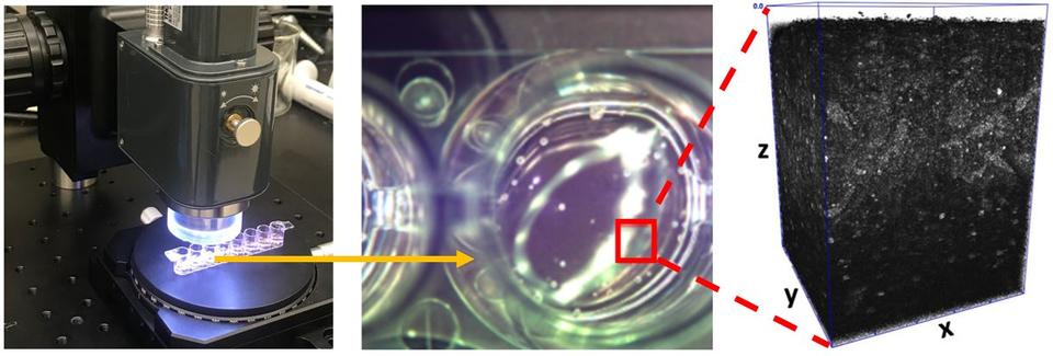
(Left) Optical coherence tomography (OCT) of samples inside an 8-well strip. (Middle) OCT brightfield camera image of the sample, where the red square encloses the observation area. (Right) 3D volumetric OCT image (1.00 mm × 1.00 mm × 1.05 mm) of the sample outlined in red (middle image).
OCT imaging was assessed using a model scaffold-cell system consisting of a polysaccharide-based hydrogel seeded with human Jurkat cells. Four test systems were used: hydrogel seeded with live cells, hydrogel seeded with heat-shocked or fixed dead cells and hydrogel without any cells. Time series OCT images demonstrated changes in the time-dependent speckle patterns due to refractive index (RI) variations within live cells that were not observed for pure hydrogel samples or hydrogels with dead cells. The changes in speckle patterns were used to generate live-cell contrast. In this way, objects with large changes in RI were binned as live cells. Additionally, the 3D distribution of live cells was mapped within a hydrogel scaffold to assess the uniformity of their distribution across the volume. Our results demonstrate a real-time, noninvasive method to rapidly assess the spatial distribution of live cells within a 3D scaffold that could be useful for assessing tissue-engineered medical products.
REFERENCES
Babakhanova G, Agrawal A, Arora D, Horenberg A, Budhathoki JB, Dunkers JP, Chalfoun J, Bajcsy P, Simon Jr CG. Three-dimensional, label-free cell viability measurements in tissue engineering scaffolds using optical coherence tomography. J Biomed Mater Res. 2023; 1- 13.
NEWS ARTICLE
Boss A. NIST Develops New Nondestructive Method for Assessing Bioengineered Artificial Tissues, 2023. https://www.nist.gov/news-events/news/2023/05/nist-develops-new-nondestructive-method-assessing-bioengineered-artificial