
Official websites use .gov
A .gov website belongs to an official government organization in the United States.
Secure .gov websites use HTTPS
A lock (
) or https:// means you’ve safely connected to the .gov website. Share sensitive information only on official, secure websites.
3D Molecular Orientation Imaging
Summary
Conventional polarization-based microscopy has been extensively used to investigate molecular alignment occurring ubiquitously in various synthetic and natural materials. However, 2D-projected anisotropy images cannot provide out-of-plane angles of molecules. Over the last ten years, the NIST Biomaterials group has developed a novel theory and experimental methods for measuring 3D orientation angles using 2D polarization imaging acquired with transmission IR and coherent Raman microscopy. This rapid, non-invasive imaging technique helps understand the molecular-level structure of highly anisotropic and spatially heterogeneous materials.
Description
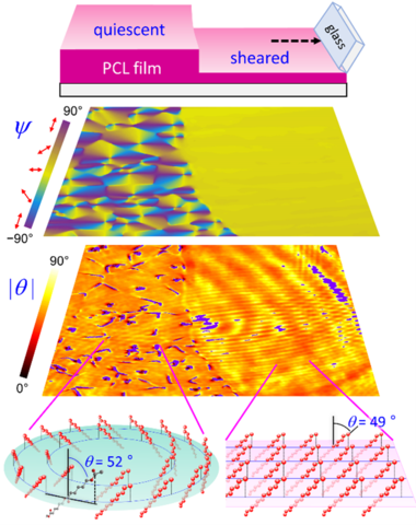
We have developed 3D orientation measurement techniques by analyzing polarization-dependent molecular vibrational signals based on infrared (IR) absorption [1,2] and coherent anti-Stokes Raman scattering (CARS) [3]. The two imaging modalities are complementary to each other: IR imaging broadly applies to any pair of non-parallel IR transition dipole moments, but its spatial resolution is in the several-micrometer scale [4,5], whereas CARS imaging can provide sub-micrometer resolution but requires more complicated data analysis [6]. These new complementary imaging methods will provide previously unavailable molecular orientation information that can bridge the gap in the relation of [process–structure–property]. This innovative optical metrology will advance plastic manufacturing, 3D printing, shape memory polymers, medical implants, tissue engineering, advanced packaging, and other material sciences and industries.
PUBLICATIONS
[1] Y. J. Lee, Concurrent Polarization IR Analysis to Determine the 3D Angles and the Order Parameter for Molecular Orientation Imaging, Opt. Express 26, 24577 (2018). https://doi.org/10.1364/OE.26.024577
[2] Y. J. Lee, Determining 3D molecular orientation from polarization-IR spectra: tutorial, J. Opt. Soc. Am. A 42, 102 (2025). https://doi.org/10.1364/JOSAA.542283
[3] Y. J. Lee, Determination of 3D Molecular Orientation by Concurrent Polarization Analysis of Multiple Raman Modes in Broadband CARS Spectroscopy, Opt. Express 23, 29279 (2015). https://doi.org/10.1364/oe.23.029279
[4] S. Xu, J. Rowlette, Y. J. Lee, Imaging 3D molecular orientation by orthogonal-pair polarization IR microscopy, Opt. Express 30, 8436 (2022). https://doi.org/10.1364/OE.449667
[5] S. Xu, C. R. Snyder, J. Rowlette, Y. J. Lee, Three-Dimensional Molecular Orientation Imaging of a Semicrystalline Polymer Film under Shear Deformation, Macromolecules 55, 2627 (2022). https://doi.org/10.1021/acs.macromol.1c02036
[6] S. Xu, Y. Jin, Y. J. Lee, 3D orientation imaging of polymer chains with polarization-controlled coherent Raman microscopy, J. Am. Chem. Soc. 144, 23030 (2022). https://doi.org/10.1021/jacs.2c10029