
Official websites use .gov
A .gov website belongs to an official government organization in the United States.
Secure .gov websites use HTTPS
A lock (
) or https:// means you’ve safely connected to the .gov website. Share sensitive information only on official, secure websites.
Summary
Quantitative chemical imaging of live cells is a long-standing measurement goal in cell-based sciences and industries. Infrared (IR) transmission microscopy is a powerful label-free imaging technique with the promise of quantitative chemical imaging. However, IR imaging of an aqueous sample remains challenging due to the strong IR absorption by water. The NIST Biomaterials Group applied the recently developed solvent absorption compensation (SAC) technique [1] to a benchtop IR microscope to eliminate the water contribution while retaining the full system dynamic range. [2] This new SAC-IR imaging system produced time traces of the total mass of three biomolecules in live fibroblast cells over twelve hours, demonstrating promise for advancing our understanding of the biomolecular processes occurring in live cells on the single-cell level. As an economical and widely adoptable imaging modality, SAC-IR microscopy could emerge as a standard imaging method of quantifying biomolecules with SI-traceability in live cells and hydrated tissues across fields such as biology, biotechnology, and medicine.
Description
The NIST Biomaterials Group introduced benchtop infrared (IR) microscopy based on a quantum cascade laser (QCL) as a quantitative chemical imaging modality of live cells. The strong IR absorption by water was mitigated by a solvent absorption compensation (SAC) technique, which pre-compensates for the strong absorption of mid-IR light by water.
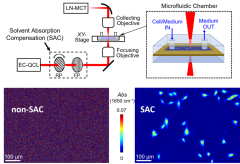
Figure 1 shows a significant reduction in absorbance noise by a two-polarizer SAC method. A series of absorbance images consisting of individual cells were analyzed to generate the absolute masses of three key biomolecules in each single cell as a function of time over twelve hours (see Figure 2). For the first time, the per-cell mass of a biomolecule species was recorded from a live cell by simply integrating the IR absorbance over a cell area. [2] The NIST team continues to improve the imaging speed and the absorption sensitivity to observe biochemical changes occurring in live cells. Monitoring the total mass of biomolecules in live cells will enable a molecular-level understanding of the metabolic processes occurring in live cells on the single-cell level.

PUBLICATIONS:
[1] B. Chon, S. Xu, Y. J. Lee, Compensation of Strong Water Absorption in Infrared Spectroscopy Reveals the Secondary Structure of Proteins in Dilute Solutions. Anal. Chem. 93, 2215 (2021). https://dx.doi.org/10.1021/acs.analchem.0c04091
[2] Y.-R. Chang, S.-M. Kim, Y. J. Lee, Benchtop IR Imaging of Live Cells: Monitoring the Total Mass of Biomolecules in Single Cells. Anal. Chem. 96, 14783 (2024). https://doi.org/10.1021/acs.analchem.4c02108