
Official websites use .gov
A .gov website belongs to an official government organization in the United States.
Secure .gov websites use HTTPS
A lock (
) or https:// means you’ve safely connected to the .gov website. Share sensitive information only on official, secure websites.
Bioimaging—A 21st-Century Toolbox for Medical Technology
Problem/Challenge
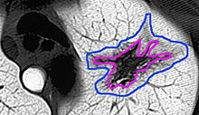
From X-ray and ultrasound images to cutting-edge 3D and 4D body images using CT scans, MRI, PET, or other advanced technologies, bioimaging is a powerful tool for viewing the internal workings of the body and its diseases.
Despite great strides in bioimaging, the field falls far short of its potential. Clinicians want to be able to treat these pictures quantitatively—to say, for example, by how much a tumor grew or shrank in response to treatment. About 40 percent of clinical trials use imaging this way.
But there is no strong measurement foundation to make those assessments accurate, reliable, and repeatable. In drug discovery, such "quantitative" imaging may be distributed among numerous test centers—and sometimes as much as 50 percent of the clinical data is useless because valid comparisons can't be made between two sites due to the lack of adequate standards.
For similar reasons, bioimaging technology usually is incapable of diagnosis—distinguishing between aggressive and benign tumors, for example. Advances in bioimaging will turn pictures into reliable data that can be used by medical professionals for diagnosis and analysis, reducing the need for biopsies and other invasive procedures and improving early detection and treatment of cancers, targeted drug therapies, and the monitoring of joint, valve, and organ replacements.
Proposed NIST Program
Many of the current roadblocks in moving from simple observation to quantitative diagnosis are issues of measurement and standards. Such a complex field requires a broad, interdisciplinary approach, marrying the physical, chemical, biological, and information sciences. NIST has strengths in all those areas and a long tradition in multidisciplinary research. NIST will partner its expertise in the physical and information sciences with the experience and know-how of the National Institutes of Health (NIH) and the bioimaging industry to develop the needed measurement capabilities.
The initiative focuses on three key areas:
- improving imaging measurement capabilities at the molecular scale (primarily through optical imaging and photonics).
- better methods (primarily based on ultrasound) to assess the behavior in the body of advanced biomaterials for cardiac repairs and treatments, angioplasty, joint replacements, and organ replacements; and
- standard methods and technologies for improved bioinformatics—capturing exchanging, combining, interpreting, and visualizing data from various imaging techniques to ensure image quality and the ability to interpret and compare data from multiple sources.
Expected Impacts
The initiative is expected to advance:
- a new generation of molecular imaging tools, allowing the identification and treatment of cancers, image-guided surgeries, and directed drug therapies;
- methods to evaluate the use of advanced biomaterials for replacing or repairing cardiac components (e.g., heart valves and stents), angioplasty, joint replacements, and organ replacements; and
- well-understood methods for image processing and analysis that will enable more reliable diagnostic imaging.