
Official websites use .gov
A .gov website belongs to an official government organization in the United States.
Secure .gov websites use HTTPS
A lock (
) or https:// means you’ve safely connected to the .gov website. Share sensitive information only on official, secure websites.
Quantitative, Traceable Determination of Cell Viability Using Absorbance Microscopy
Summary
Most cell-based protocols require knowledge of the number of live and dead cell populations. The current methods, however, lack traceability, which is particularly important in biomanufacturing. Quantitative and traceable measurements of cell viability are greatly needed to improve consistency in the biomanufacturing of cells and cell-based therapeutics. This work demonstrates a new imaging modality, absorbance microscopy, which was developed for traceable cell viability measurements. It enables the collection of comparable images of cells over time on the same microscope or on different microscopes.
Description
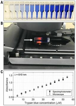
The current gold standard for cell viability measurements is the trypan blue assay. Trypan blue dye enters dead cells that have a ruptured membrane. As a result, the dead cells are stained blue. Live cells with an intact membrane, on the other hand, do not absorb the dye molecules and remain unstained. Thus, when live and dead cells are mixed with trypan blue solution, one may count the stained and unstained cells to determine the number of live/dead cells as well as the percent viability of that sample. Such an experiment is typically performed using a brightfield microscope, where the intensity of the pixel values is utilized to determine whether the cell is light (unstained/live) or dark (stained/dead). Such intensity measurements of the sample are not suitable when comparing measurements performed on the same microscope at another time or on a different microscope. Variations in optics and microscope cameras may alter the intensity values for the same sample.
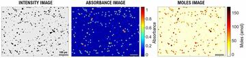
In this work, quantitative and traceable live-dead cell classifications are performed by calculating the amount of trypan blue dye present in single cells. The intensity pixels of the brightfield images are converted to absorbance images that are used to calculate moles of trypan blue per cell (see the figure above). To implement the absorbance microscopy method, we developed an open-source AbsorbanceQ application that generates quantitative absorbance images. Instead of making live-dead classifications using arbitrary intensity values, absorbance imaging yields traceable units of moles that can be compared, which is useful for assuring quality for biomanufacturing processes.
Major Accomplishments
Greta Babakhanova, Stephen M. Zimmerman, Laura T. Pierce, Sumona Sarkar, Nicholas J. Schaub, Carl G. Simon Jr.
Quantitative, Traceable Determination of Cell Viability Using Absorbance Microscopy. PLoS ONE 17 (1), e0262119, (2022).
https://doi.org/10.1371/journal.pone.0262119
Greta Babakhanova, Stephen M. Zimmerman, Laura T. Pierce, Sumona Sarkar, Nicholas J. Schaub, Carl G. Simon Jr.
Dataset for Absorbance Microscopy for Quantitative and Traceable Trypan Blue Cell Viability Measurement
https://datapub.nist.gov/od/id/mds2-2347
Stephen M. Zimmerman, Carl G. Simon, Jr., Greta Babakhanova
AbsorbanceQ: An App for Generating Absorbance Images from Brightfield Images. J Res Natl Inst Stan 126:126039, (2021).
https://doi.org/10.6028/jres.126.039
Stephen M. Zimmerman, Carl G. Simon, Jr., Greta Babakhanova
AbsorbanceQ App for Generating Absorbance Images from Brightfield Image Captures
https://data.nist.gov/od/id/mds2-2423