
Official websites use .gov
A .gov website belongs to an official government organization in the United States.
Secure .gov websites use HTTPS
A lock (
) or https:// means you’ve safely connected to the .gov website. Share sensitive information only on official, secure websites.
How Does an MRI Machine Work?
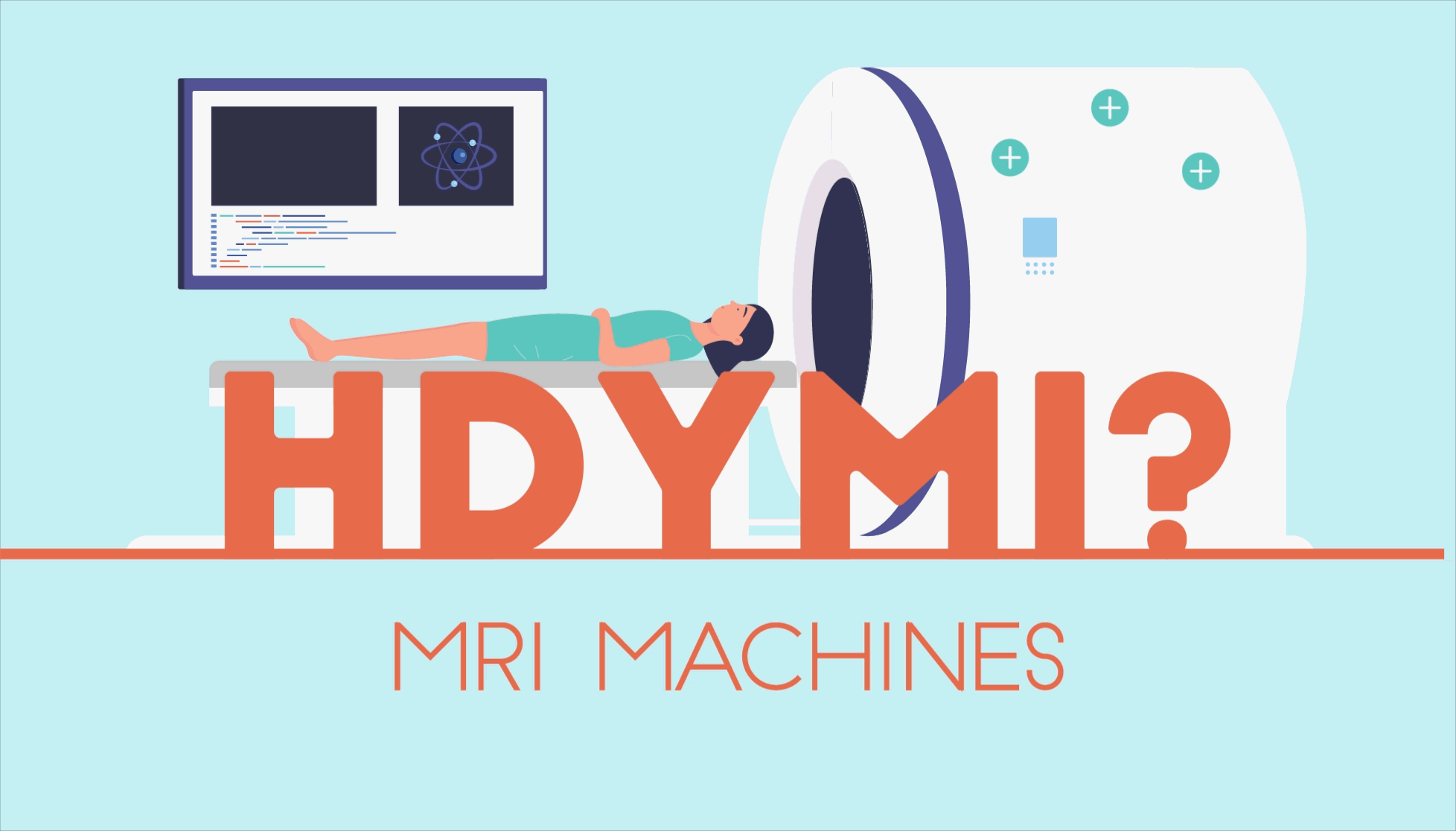
MRI stands for magnetic resonance imaging. If you’ve ever had an MRI, you may wonder how this machine can “see” inside your body. The answer lies at the subatomic level.
Protons are small, positively charged particles found in atoms. In addition to having an electric charge, protons have tiny magnets, so they interact strongly with magnetic fields. Humans have many, many protons. The protons are mostly from the water in our bodies, but also from other molecules such as proteins and fat.
An MRI machine contains a magnetic field, hence the “magnetic” in its name. When you enter the machine and the magnetic field turns on, the protons in your body respond strongly to this field. You can imagine each proton having a bar magnet pointing through it. The MRI system’s magnetic field lines up some of the bar magnets, so they are all pointing in the same direction.
The MRI machine then uses a radiofrequency pulse of invisible light to tip the protons’ bar magnets out of this alignment. For the tipping to work correctly, this radio wave must have a particular frequency, which depends on the basic properties of the protons. This frequency matching is the “resonance” in MRI. If you’ve ever heard the phrase “on the same wavelength,” this is a similar concept!
After the radiofrequency pulse passes through the body, the protons’ little magnets move back into their original positions. As the protons “relax” back into these positions, they release electromagnetic signals that the MRI machine “sees.” These signals contain information about the protons’ position in the body as well as their environment (such as healthy tissue or tumors).
We’re not done yet, though. The MRI machine needs to repeat the process of firing radiofrequency pulses many times to build up images. Each time, the machine turns on spatially varying magnetic fields (known as magnetic field gradients) to encode the location of protons within the MRI signal. The spatial information is critical for creating a 3D image.
When the scan is finished, computers combine information from all the signals to create images of the body. These are the images that doctors use for diagnostic purposes.
A New Frontier in MRI Technology
MRIs were invented in the 1970s and have become an important diagnostic tool. In fact, an estimated 40 million MRIs happen each year in the U.S.
MRIs provide qualitative information — pictures of the inside of the body.
But what if an MRI could provide “quantitative” information in the form of measurements? This is the goal of a research effort here at NIST — to build toward quantitative MRI that provides data and measurements, not just pictures.
Quantitative MRIs are currently used in clinical trials for medical therapy and in some clinical settings. Researchers conducting clinical trials on cancer treatments, for example, need to be able to measure patients’ tumors over time. They need to know that if a tumor is shrinking, it’s because the medication is working and not because of variances in the measurements given by different MRI machines examining individual participants.
That’s where a reference object known as a “phantom” comes in. A phantom made at NIST allows MRI machines to be calibrated to make sure they provide accurate, quantitative measurements.
Doctors use quantitative MRIs in specialized applications now, such as treating liver disease. With additional research, quantitative MRIs have tremendous potential to better diagnose diseases and test new treatments.
As this vital research continues, NIST researchers are working to learn everything we can about MRIs and how they can take the most accurate measurements possible. Researchers are collecting data and trying to answer critical questions, such as:
- How often do you have to take measurements to ensure accuracy?
- What changes happen to an MRI machine over time, and how do we account for those changes in measurements?
Mobile MRIs Can Make Health Care More Accessible
MRIs are typically located in hospitals or medical centers because the machines are large, and people and equipment are needed to operate them. But new technology is shrinking the MRI machine, and some machines can now travel to the patient.
Thanks to recent advances, some MRI machines are small enough to fit in the back of a truck or to be brought into an ICU and plugged into a simple wall outlet, though they are not yet in widespread use. Known as low-field MRI, these machines have the potential to make health care much more accessible to people who don’t have a hospital nearby.
One of the challenges with low-field MRI is the quality of the images, but we’re also studying how to use AI to improve these images and demonstrate the clinical utility of these systems. The goal of this work is to make low-field MRIs more useful as they become more common.
More research is needed to make sure these machines take the most accurate possible measurements. But measurement is what NIST does. We’ll keep learning more about these new developments in MRI technology — so you can be your healthiest.