
Official websites use .gov
A .gov website belongs to an official government organization in the United States.
Secure .gov websites use HTTPS
A lock (
) or https:// means you’ve safely connected to the .gov website. Share sensitive information only on official, secure websites.
Taking Measure
Just a Standard Blog
At some point in my teenage years, I became aware that when I grew up, I’d have to get a mammogram, and it was clear that it would hurt.
If you’re a woman over 40 (or you’ve talked to women over 40 about this topic), you probably know that this annual screening requirement for all women is uncomfortable at best and can be painful. Can you imagine, a woman who lives into her 80s may have to undergo this test 40 or more times throughout her life?!
But it is a lifesaving test that all women (and some men) need.
Now, I’m a medical imaging researcher, and I know that a less painful alternative to breast cancer screening is possible within my lifetime.
I have a goal some might consider outrageous — to create a viable alternative to mammograms that is pain-free, highly accurate and can save even more lives than mammograms currently do.
Challenges of Mammography
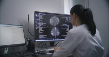
Mammograms use low-dose X-rays to take a picture of the inside of the breast. The breast must be compressed to increase the image quality, but that compression makes it uncomfortable.
Mammograms are incredibly important tests that have saved many lives. Right now, they are the best option we have to detect and treat breast cancer early, and everyone eligible for mammograms should prioritize getting them.
But that doesn’t mean there aren’t drawbacks.
First, mammograms can miss up to 35% of breast cancers.
On the other end of the spectrum, mammograms also can give false positives up to 14.4% of the time. This can mean extra tests, including painful biopsies, for patients to rule out breast cancer.
This risk is especially high for the up to half of women over age 40 who have dense breast tissue. Breasts are made up of two types of tissue — dense tissue and fatty tissue. Women with dense breasts have more dense tissue. Dense tissue is normal and nothing to be concerned about, but dense tissue makes mammography more challenging and can lead to false positives.
Low-Field MRI and Breast Cancer Screening
My research focuses on lower-strength magnetic-field equipment, known as low-field MRI. Unlike a traditional MRI, which is a large, expensive machine, low-field MRI machines are smaller and less costly. That’s because they use a weaker magnetic field to capture an image. The weaker magnetic field means the images are not as clear as traditional MRI.
My goal is to use this same technology to create a way to screen patients for breast cancer. Breast imaging with traditional MRI can be done now, and it does not require the breast compression used for mammography. But MRI is expensive and is often reserved for patients who need follow-up testing. With low-field MRI, this option could be available to many more patients.
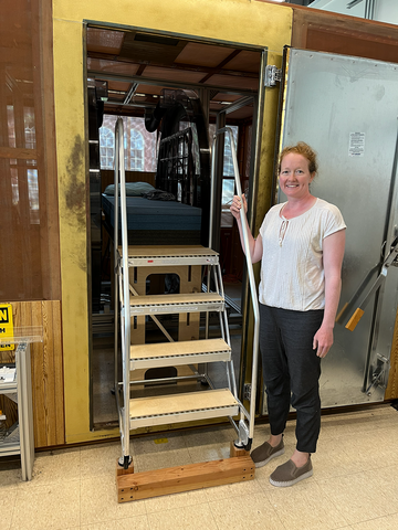
I worked with imaging researchers Matthew Rosen, Neha Koonjoo and Sheng Shen and Dr. Mansi Saksena and colleagues in Boston to modify their existing ultralow-field MRI machine for breast imaging, and we recently tested it with a small group of patients. The machine costs a few hundred thousand dollars to build, a fraction of the several million dollars that a typical MRI system costs.
During its development, I tried out the system, and I found it to be a painless experience. I couldn’t say the same about the mammogram I got a few months later!
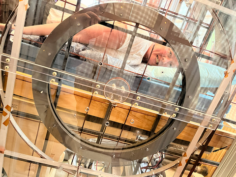
In the initial study, we used the low-field MRI machine to image one breast each from a few women with no history of breast cancer and a few with breast cancer. Obviously, for this technology to work, we need to be able to image both breasts at once. But for this study, we started with just one at a time.
One important caveat is that our machine has not yet actually detected breast cancer. That means we still have a lot of work to do to get this technology to a point where it could replace a mammogram, but we’re in an exciting, exploratory phase of this research.
In other ways, our study was successful. The radiologists were impressed with how much they could see in the low-field MRI images. You can view images throughout the breast, which you don’t typically see on a mammogram. Being able to see more could mean that a low-field MRI could detect breast cancer at a higher success rate than traditional mammograms.
To me, many body parts appear similar from person to person on an MRI or X-ray. I used to work in knee-related research, and all knees pretty much look the same on the inside. So far, every breast looks different on MRI, and I think that’s beautiful and interesting. I hope our research can one day help give radiologists the best possible imagery to diagnose breast cancer.
Better Screening Will Better Serve Women
I hope that with continued time and research, low-field MRI will make breast cancer screening less painful for patients — and possibly less costly overall for the medical system.
One possibility is that this technology could be integrated into the surgical suite where doctors remove breast cancer from patients. One of the frustrating aspects of breast cancer treatment is that the surgeon doesn’t always know right away that they took out all of the cancer during surgery.
If low-field MRIs become readily available, these machines could be used right in surgical suites. A doctor could theoretically check to make sure they’ve gotten the entire cancerous mass out before bringing the patient out of anesthesia, reducing the chances that a patient would have to undergo multiple surgeries.
Our team is continuing to experiment with different magnetic field strengths to find the best image. We’re also looking at the most optimal way to be able to image both breasts at once, which will be an important part of this process as we advance.
This is a gratifying project for me to work on as a researcher, both as a woman with dense breasts who has had to have follow-up testing done, and as the friend and relative of many women who’ve had breast cancer.
A woman in the U.S. has a one in eight chance of developing breast cancer. That’s a scary number, but the good news is that more women are being diagnosed early and surviving than ever before.
We have a long way to go before low-field MRI becomes a common alternative to mammograms. But my fellow researchers and I will continue to work to make this happen, because all patients deserve a highly accurate and pain-free approach to screening for breast cancer.
About the author

Related Posts


Comments
Thank you for working on this!! It is much needed!
I always wondered why a better way hasn't been invented yet, so great work here! I think this method might also be less scary for women so it encourages them to get screened earlier and more often!
I’m a breast cancer survivor. I have dense tissue, so the recommended follow up is alternating mammograms and MRIs. But I can’t have MRIs due to an implanted device, so I have ultrasound. I’d love a less painful alternative and I was actually wondering if anyone was looking for one after my last mammogram. I’m really glad someone is, even if it might not help me specifically. It’s long overdue
I applaud this work and greatly hope that it enables more accurate AND more comfortable breast cancer screening!