
Official websites use .gov
A .gov website belongs to an official government organization in the United States.
Secure .gov websites use HTTPS
A lock (
) or https:// means you’ve safely connected to the .gov website. Share sensitive information only on official, secure websites.
Taking Measure
Just a Standard Blog
Giving radiation to a cancer patient requires precise measurements. You need to give a lethal dose to the tumor and a nontoxic dose to the surrounding tissue. NIST researchers are working to improve these measurements, helping people with cancer live healthier lives after they complete their treatment.
In the evenings, after patients have left for the day, our research team visits the radiation oncology offices at the University of Colorado Anschutz Medical Campus to talk to medical physicists about how our research can help cancer patients. We also run experiments in their radiation suites.
The radiation suites contain large machines that deliver high-energy X-ray and electron beams, known as accelerators. The rooms are surrounded by foot-thick concrete walls with sliding doors. The patient beds and accelerator heads move around in complex and precise motions to deliver radiation uniquely tailored to each patient and tumor.
We arrive after the medical physicists have just finished a busy day. The medical physicists test and calibrate the radiation systems. They also prepare radiation dose plans based on imaging and complex patient-specific calculations. Dose planning is a multistep procedure that requires accuracy and precision to ensure that the person gets the exact amount of radiation needed. You want to give a lethal dose to the tumor and a nontoxic dose to the surrounding tissue.
It’s a sobering reminder of the importance of our work — developing methods to better measure and image radiation. Our goal is to help cancer patients live long, healthy lives for decades after they complete treatment.
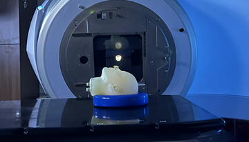
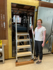
Back in our lab at NIST, we’re creating a human-head-shaped container filled with radiation-sensitive material. It’s called an anthropomorphic dosimeter. Anthropomorphic means it looks like a person, and dosimeter means it measures radiation.
The head replica, which we call a phantom, contains a special radiation-sensitive gel. The gel is mostly water, and it mimics tissue. The phantom also contains special polymers that change their chemistry when exposed to radiation.
The phantom can be thought of as a three-dimensional radiation-sensitive photographic film. When radiation hits parts of the phantom, we can record changes in the film. We do this using a special type of MRI that provides data and measurements, not just pictures, known as quantitative MRI. It is our expertise in quantitative MRI that brings this new opportunity to radiation dosimetry.
Using the quantitative MRI rather than a traditional MRI, we can better measure where and how much radiation is deposited and use this information to verify the accuracy of dose plans. We’re planning to use these gels to study different types of radiation, such as X-ray, electron, proton, ultrasound and microwaves. We hope to better understand how and where they are absorbed in the body.
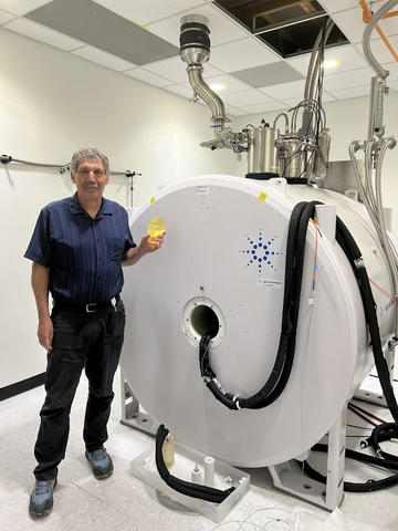
The long-term goal is to produce a cost-effective phantom and tabletop MRI system that can go into radiation oncology suites and help patients where they’re receiving care to get precise, personalized treatment.
Luckily, getting measurements just right is what we do here at NIST.
This research is gratifying because people are living for a very long time after cancer treatment, sometimes 20-30 years or more. We need to understand the long-term effects of radiation and minimize the side effects so people can live healthy, productive lives.
One study showed that the long-term cost of health care after radiation can often exceed the actual cancer treatment expenses, so there’s a financial benefit, in addition to improving people’s quality of life.
Magnetism and MRIs
I have worked in magnetism and related research for the last 35 years. I got interested in MRI because it uses magnetic fields to probe the human body. Nature has provided every electron and proton with a magnetic property called spin that sends out radio waves. The frequency of the waves can sense the location and activity of biomolecules such as water. MRI machines take advantage of spin to take images of the body.
In 2005, medical societies, pharmaceutical companies and other stakeholders asked NIST to lead a program on standards for quantitative medical imaging. I stepped up to the challenge and created the NIST MRI standards program, which was funded by Congress in 2007.
Our work on radiation dosimetry started in 2020 when we realized that our quantitative MRI techniques could be used to image, in 3D, the changes in specially sensitized materials that mimic tissue during radiation. NIST began collaborations with international groups working on MRI-readable radiation dosimetry, and we’ve been working to advance this science since then.
I have been motivated by personal experience. My mother was treated at age 65 for an abdominal tumor. We are grateful that surgery and radiation led her to be cancer-free at the age of 90, but collateral damage led to many years of health challenges. I wish things could have been measured and applied more precisely.
Looking Ahead to Future Health Care Measurements
One of the most exciting aspects of this research is that once it's perfected, we can expand it to other measurements beyond dosing. One area is in our DNA. Our DNA is always getting damaged and repairing itself; that’s a normal part of life. Excessive radiation can disrupt this process, and irreparable DNA damage can lead to cell death and cancer. We’re working to sensitize our gels to measure DNA strand breakage, so we can study this damage.
Our measurements and research have also been used to improve small, portable MRIs, which have increasingly become available to health care providers. This technology helps make MRIs more accessible to people in ambulances, mobile settings or areas without easy access to hospitals.
In addition to these health care examples, we have developed a method to image cellphone radiation based on quantum sensing — an important topic for us to study, given the ubiquity of these devices in nearly everyone’s life.
We’re also looking to measure radiation exposure in space. That’s because if humans are going to spend any amount of time in space, we need to know how to do so in a healthy way. Part of that will be studying the amount of radiation in space and understanding our bodies’ ability to tolerate it.
NIST physicists have a lot to contribute to improving our health care system. Measuring things precisely and measuring things we could not see before will improve treatment quality, reduce costs and provide better well-being for ourselves, our relatives and our society.
How Do MRIs Work?
Ever wonder how an MRI machine gets a glimpse inside your body? Learn more in our How Do You Measure It? explanation.
About the author
Related Posts

Comments
Have you talked with the ionizing radiation division in Gaithersburg, MD - Dr. Michael Mitch. They have all the standards used for cancer treatments with radiation therapy.
Thank you for your comment. We work with the NIST ionizing radiation group in Gaithersburg and have submitted some joint proposals to merge MRI dosimetry with SI-traceable dosimetry. We will also be working with the Medical Physics Division at NPL in London. One interesting distinction is that we image both ionizing and nonionizing radiation. We also work on measuring other parameters, to complement dose, to provide additional information on the interaction of radiation with tissue. If you are interested in this work, please come and see us at the American Association of Physicists in Medicine (AAPM) conference at the end of this month in Washington DC.
Thank you Dr. Stephen Russek and NIST for sharing this interesting and important work!
Grateful for the incredible work NIST is doing to push the boundaries of healthcare, from improving access to portable MRI technology, to helping us understand how radiation exposure affects the human body in space.
Standardizing these kinds of measurements isn't just technical. It’s essential for building safer, more equitable, and future-ready healthcare systems. Excited to see this kind of innovation making real-world impact! Great work Dr. Stephen Russek.
This is an excellent article on really impactful work. Thank you, Stephen and team!