
Official websites use .gov
A .gov website belongs to an official government organization in the United States.
Secure .gov websites use HTTPS
A lock (
) or https:// means you’ve safely connected to the .gov website. Share sensitive information only on official, secure websites.
Microresonators for inductive-detection electron paramagnetic resonance spectroscopy of volume-limited samples
Summary
Planar microresonators are fabricated in the NIST Nanofab, tested in our laboratory, and incorporated into a commercial 9 GHz or NIST-built 34 GHz electron paramagnetic resonance (EPR) spectrometer for measurements of thin films and other volume-limited samples. The sensitivity of our microresonators is predicted to be at least two orders of magnitude higher than conventional cavity resonators. The combination of higher sensitivity and reduced size of the microresonators enables measurement of EPR spectra of samples such as monolayer/multilayer thin films and interfaces. Interrogation of volume-limited soft matter samples, for example, biomacromolecules and polymers, also is being pursued.
Description
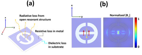
Figure 1: (a) Schematic of the planar inverse anapole resonator and the types of power losses occurring from planar microresonators. The purple shaded region is metallized while the transparent cuboid is the dielectric substrate on which the metal layer is deposited. (b) Left panel - red arrows indicate the electric dipole moment while the yellow arrow indicates the toroidal moment in a planar inverse anapole resonator. Mutual cancellation of the electric dipole moment and toroidal moment leads to diminished far-field radiative losses. Right panel - active region of the resonator, which is the metallic bridge at the center of the structure. The microwave excitation field B1 is confined to a sub-wavelength scale (nL to pL volume) around the central bridge.
Spin-detection in volume-limited samples has applications ranging from solid-state physics to structural biology. Increasingly, technologically-relevant materials have nanoscale dimensions. Single microcrystals may only have volumes ranging from picoliters to nanoliters. Many biological materials, particularly membrane-embedded proteins, are laborious to obtain in sufficient volume and concentration for conventional structural biology analysis methods. Magnetic resonance spectroscopies based on inductive detection are powerful and versatile techniques that can provide atomic-level structural and functional information about a wide range of samples under broadly variable conditions. These methods, however, are inapplicable for nanoscale samples; for example, inductive-detection electron paramagnetic resonance (EPR) spectroscopy currently requires sample volumes on the order of tens of microliters.
A popular and successful way of decreasing detection limits is to miniaturize the volume being interrogated by creating resonators from planar structures patterned on dielectric substrates. These microresonators can be readily fabricated using standard photolithographic techniques, which provide micrometer-scale dimensions resulting in nanoliter active volumes. When the active volume becomes comparable to the sample volume, there is a gain of in absolute sensitivity. Miniaturization inevitably deteriorates the ability of the resonator to retain absorbed energy, which adversely impacts overall sensitivity gains. Compared to cavity resonators, power losses from resistive, dielectric, and radiative losses (Figure 1a) are greater in open resonant structures such as loop-gap resonators and microstrip-based structures. Our microresonators target radiative loss (Figure 1b) to increase the microresonator sensitivity in a picoliter active volume.
Publications
Scalable microresonators for room-temperature detection of electron spin resonance from dilute, sub-nanoliter volume solids by N. Abhyankar, A. Agrawal, P. Shrestha, R. Maier, R.D. McMichael, J. Campbell, V.A. Szalai, Science Advances 28 Oct 2020 Vol. 6, no. 44, eabb0620. DOI: 10.1126/sciadv.abb0620.