
Official websites use .gov
A .gov website belongs to an official government organization in the United States.
Secure .gov websites use HTTPS
A lock (
) or https:// means you’ve safely connected to the .gov website. Share sensitive information only on official, secure websites.
Summary
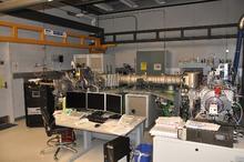
Magnetic sector secondary ion mass spectrometry (SIMS) generates isotopic and elemental information from solid surfaces with depth profiling capabilities to measure the concentration of isotopes and elements as a function of depth into the film.
Description
Magnetic sector SIMS uses high energy “primary” ions to bombard the surface and liberate “secondary” ions much like in ToF-SIMS, but the ion beam is operated continuously to maximize duty cycle. The high ion dose is preferred for depth profiling, where the concentration of elements and isotopes are determined as a function of depth. This is one of the very few techniques where the depth resolution can approach 1 nm in inorganic films. The separation of “secondary” ions by mass occurs through a double-focusing lens, first through an electrostatic sector to filter the ions by energy, then through a magnetic sector to filter the ions by mass. Quantification is achieved by applying relative sensitivity factors (determined from standard samples) to convert secondary ion intensities to concentrations. Precision and accuracy better than 1 % can be achieved with detection limits in the parts-per-million (ppm) to parts-per-billion (ppb) for most elements in the periodic table.

Current Activities
Method and Standards Development for Improved Isotopic Measurements
SIMS is used to characterize the elemental and isotopic compositions of solid materials ranging in size from sub-µm to 100s of µm in fields including semiconductors, cosmo/geochemistry, and nuclear materials. One of the SIMS capabilities in the Surface and Trace Chemical Analysis Group is the Large-Geometry (LG)-SIMS, which can achieve high transmission at high mass resolving power, and represents the state-of-the-art for SIMS sensitivity and selectivity, particularly for trace analytes. Like many mass spectrometry techniques, SIMS is a highly precise method for relative quantification, in that it relies on “ground-truth” standards to yield absolute isotopic and elemental abundance information.
One field of international importance is Nuclear Safeguards, which relies on the ability to precisely measure the composition of actinide particles sampled at U.N. Member States’ nuclear facilities. Appropriate particle standards are required for quality control and bias correction of these analyses. We work with partners to help develop and characterize new particle standards tailored for specific SIMS analyses and develop new measurement paradigms that improve the sensitivity for ultra-trace isotopic analyses of these particles. Two recent developments from our group include a uranium particle age dating method using the 234U→230Th radiochronometer and the use of new primary ion species (i.e., O3-) to enhance the measured signal of U and Th by 100 % relative to conventionally used O-.
SIMS is also commonly applied to the quantification of shallow ion implants and dopants in semiconductors and other materials. We are working on characterizing the elemental and isotopic composition of the solar wind collected by NASA’s Genesis Discovery Mission and returned to Earth. The composition of the solar wind reflects that of the Sun (nearly 99.9 % of the mass of the solar system) and is used to model the mechanisms of solar wind acceleration and enhance space weather prediction. This is crucial for protecting satellites, which we all rely on heavily for communication, commerce, navigation, and much more.
Publications
"Improved uranium particle analysis by SIMS using O3− primary ions", Groopman, Evan E., Williamson, Todd L. and Simons, David S., J. Anal. At. Spectrom., (2022) 37 2089-2102. http://dx.doi.org/10.1039/D2JA00231K.
"Multi-collector Configuration Considerations And Substrate Relative Sensitivity Factor Effects For Age-dating Measurements Of Particles By Large Geometry Secondary Ion Mass Spectrometry", Todd L Williamson, David S Simons, John D Fassett, Proceedings of the INMM & ESARDA Joint Virtual Annual Meeting August 23-26 & August 30-September 1, 2021. Abstract