
Official websites use .gov
A .gov website belongs to an official government organization in the United States.
Secure .gov websites use HTTPS
A lock (
) or https:// means you’ve safely connected to the .gov website. Share sensitive information only on official, secure websites.
2D and 3D image Measurements of Retinal Pigment Epithelial Cells at Terabyte Scale
Summary
Progressive increases in the number of cell therapies in the preclinical and clinical phases has prompted the need for reliable and non-invasive assays to validate transplant function in clinical biomanufacturing. This general need has motivated our work to deliver trusted measurements and quality metrics for tissue-engineered implants, such as the implants consisting of retinal pigment epithelium (RPE) cells for treating vision loss in adults. We focus on microscopic image-based 2D and 3D measurements of RPE cells that have been stained with tens of biomarkers and imaged over 6 weeks at multiple fields of view and with a variety of treatments. Such 2D and 3D image collections reach several terabytes in size and present a challenge for deriving trusted measurements.
Description
Our objectives include:
- the design of robust 2D and 3D measurement platforms for characterizing RPE cell implants and
- the demonstration of such measurements from bright-field microscope and fluorescently labeled images to predict the biological function of an RPE tissue.
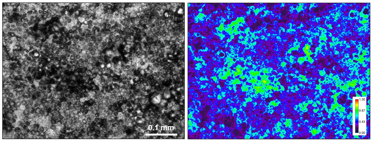
The 2D measurement platform is composed of quantitative bright-field absorbance microscopy (QBAM) and deep neural networks (DNNs) to non-invasively predict tissue function and cellular donor identity. The 3D measurement platform is based on fluorescent confocal microscopy, model-based analyses, and multi-channel (stain) quantifications of cell organelles to assess tissue quality. Figure 1 illustrates images of bright-field and quantitative bright-field absorbance images.
Major Accomplishments
QBAM images of iRPEs were used to train Deep Neural Networks (DNNs) that predicted iRPE monolayer transepithelial resistance (TER) (R2=0.97), predicted polarized Vascular Endothelial Growth Factor (VEGF) secretion (R2=0.89), and matched iRPE monolayers to the iPSC donors (accuracy=85%). DNN predictions were supplemented with traditional machine learning algorithms that identified shape and texture features of single cells that were used to predict tissue function and iPSC donor identity. These results demonstrate non-invasive cell therapy characterization can be achieved with quantitative bright-field microscopy and machine learning. For details, see the publications below.
Associated Product(s)
Resources/Demos
Data dissemination and interactive visualization
A DOI has been minted for the data record: 10.18434/T4/1503229