
Official websites use .gov
A .gov website belongs to an official government organization in the United States.
Secure .gov websites use HTTPS
A lock (
) or https:// means you’ve safely connected to the .gov website. Share sensitive information only on official, secure websites.
Automated Live Cell Imaging for Quantification of Catalytic Rates in Individual Cells
Summary
To accurately interpret the data from fluorescent proteins as reporters of gene activation within living cells, it is important to understand the kinetics of the degradation of the reporter proteins. However, quantitative measurements of dynamic processes in single, living cells are challenging, especially when the processes being studied are long-lived and the number of cells required to achieve an accurate determination of the distribution of responses in the population is large. In this study, the rates at which large numbers (>500) of individual living cells enzymatically degrade green fluorescent protein (GFP) were examined using simple protein patterning techniques and automated fluorescence microscopy.
Description
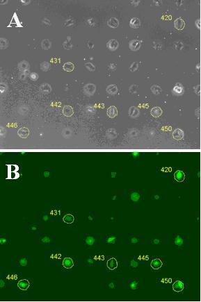
In this study, the rates at which large numbers of individual living cells enzymatically degrade green fluorescent protein (GFP) was examined using automated fluorescence microscopy. A clonal population of NIH3T3 fibroblasts that resulted from stable transfection with a construct containing the tenascin-C gene promoter ligated to a destabilized, enhanced GFP (dsEGFP) reporter gene was studied. We used simple protein patterning methods based on soft microlithography to confine cells to discrete spatial locations coated with the extracellular matrix protein, fibronectin. Spatial confinement facilitated the analysis of GFP degradation rates by limiting the migration of the live cells during the 16 hours of the experiment. Cells continued to move within and slightly beyond the designated areas during the experiment. To generate a segmentation mask for cells which included the entire area that each cell explored, sequential phase images were subtracted, and the absolute difference in intensities of each pixel in the images were summed. Pixels above a manually set threshold provided a mask under which the GFP intensity for individual cells was determined (see Figure).
This method allowed us to examine GFP intensity over 14 h in 524 live cells and determine the degradation rate constants for GFP in those cells. To measure GFP degradation rates in the absence of new GFP production, protein synthesis was inhibited by treating the cells with cycloheximide. Results indicated a wide range of cell-to-cell variability in the GFP fluorescence within individual cells, and a non-Gaussian distribution of intensities. Cell-to-cell variability in degradation kinetics was relatively narrow, and more symmetrical. Degradation for this reporter was analyzed as a first order rate process with a degradation half-life of 2.8 +/- 0.7 h, where the variance reflects the width of the distribution of rate constants.
By examining degradation rate as a function of initial GFP intensity, we found that GFP degradation rate constants were independent of the initial intensity of GFP fluorescence within cells. This result suggests that higher GFP abundance in some cells is likely due to higher rates of gene expression, because it is not due to systematically lower rates of protein degradation.
Major Accomplishments
- Measured GFP intensities from live, single cells for over 14 hours using simple protein patterning methods and automated microscopy
- Determined the distribution of degradation from 499 of those cells and found that the observed GFP intensity within a cell is independent of the degradation activity within that cell
- Found that the distribution of rate constants for enzymatic degradation of this protein in cells is narrow compared to other cellular phenotypic parameters.
Additional Technical Details
M. Halter, A. Tona, K. Bhadriraju, A. L. Plant, J. T. Elliott, "Automated live cell imaging of green fluorescent protein degradation in individual fibroblasts." Cytometry A. 2007 Oct;71(10):827-34.