
Official websites use .gov
A .gov website belongs to an official government organization in the United States.
Secure .gov websites use HTTPS
A lock (
) or https:// means you’ve safely connected to the .gov website. Share sensitive information only on official, secure websites.
Researchers at the National Institute of Standards and Technology (NIST) and colleagues have developed a novel, anatomically accurate reference model of the human brain, entirely made from soft organic materials, for use in magnetic resonance imaging (MRI).
The work revealed several major obstacles to fabricating anthropomorphic phantoms, including the persistent presence of “signal voids” – blank areas in the MRI images caused by the boundaries between different kinds of tissue mimics. And an innovative technique to mimic cerebral “microbleeds” that often accompany traumatic brain injuries (TBI) was not fully successful.
The team offers detailed descriptions of the now completed project, along with suggestions for alternative techniques, in the July 12, 2023 issue of PLOS ONE to encourage other groups to build on their experience.
Artificial reference objects that, when imaged, mimic the properties of real tissue – and thus can be used to calibrate MRI and other imaging systems for medical diagnosis and treatment – are called phantoms. NIST has pioneered numerous MRI phantoms, such as the recent breast phantom design that was rapidly and widely adopted. And it has been a world leader in “quantitative” MRI – which measures the types and intensities of signals from tissue – as opposed to “qualitative” approaches that chiefly rely on image contrast between adjacent areas.
But brain phantoms pose numerous special challenges, and to date “no 3D anthropomorphic brain structure suitable for whole-brain quantitative MRI has been developed,” the authors note. The NIST team, including partners from Mitre Corp., Hyperfine Inc., and the University of Colorado, has been at work on the problem since 2017, with interruptions resulting from the COVID pandemic.
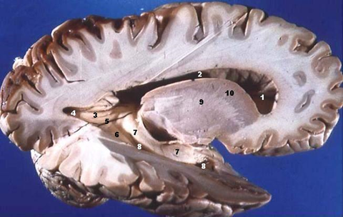
“In the brain we have gray matter and white matter, and they are seamless,” said NIST lead researcher Katy Keenan. “But constructing something that is seamless is very difficult. Conventional brain phantoms typically have rigid materials between different types of tissue mimics, and those show up on the scanner as dark artifacts, very obviously different from how the actual brain appears.
“So what we’ve been trying to do is come up with something that doesn’t have those boundaries that produce signal voids.”
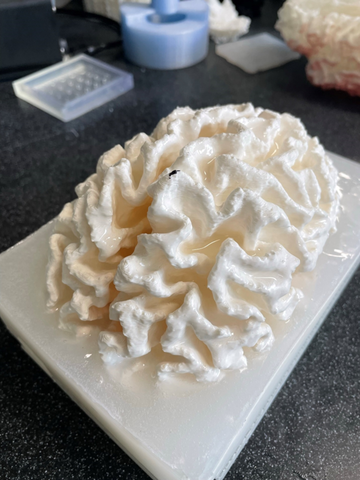
The researchers worked with frameless molded shapes of both white and gray matter, including the characteristic folds and fissures. Gray matter occupies the outermost regions of the brain and contains neural cell bodies and the synapses by which cells interact. White matter consists of bundled axons that process and route nerve signals to the spinal cord. For the NIST team’s phantom, the two tissue types were made from differently doped gels chemically related to the nutrient in petri dishes familiar from high school biology.
The NIST team used a dataset available from The Martinos Center at Harvard University and Massachusetts General Hospital for their white matter mold and outer skull.
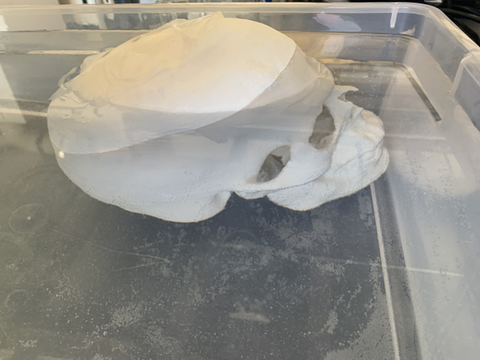
They used various molding methods, including a hard cast for the white matter that was employed to shape the gel as it solidified. Then the cast was dissolved from around it. Both kinds of soft materials were coated with a waterproof spray and placed in a 3D-printed solid skull. None of the methods produced images that were entirely artifact-free.
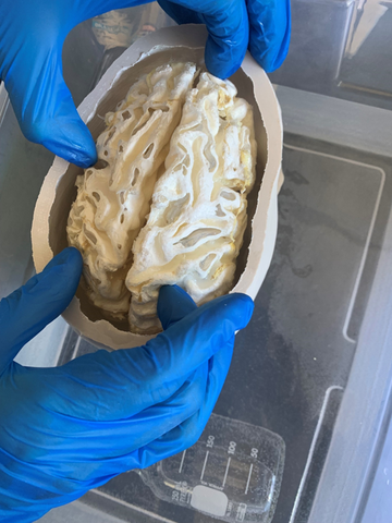
Before deciding to use the molding methods, the team tried several approaches. They considered using a mold like a silicone ice cube tray, which they were hoping to peel off the gel. But the folds in the white matter were problematic and some of the gel broke off with the mold. They also considered 3D printing the two gels directly, but that wasn’t possible at the size of the brain. Dissolution of the 3D-printed cast worked well on small test pieces. However, it did not work as planned at the scale of the white matter. The mold around the white matter wasn’t fully dissolved. ( See photo.)
While the team was disappointed that this project was only moderately successful, they are excited to see how another group might solve the problem. The team hopes that by making this research available another group will learn from their lessons and have success with an approach that builds on this work.
“A fully 3D anthropomorphic phantom enables us to test MRI scanners in a way that we currently cannot,“ said NIST researcher Stephen Russek. “Using a brain-like shape can reveal issues with the scanner that we miss with conventional, geometric phantoms.”
Paper: Mikail Kraft, Slavka Ryger, Ben P. Berman, Matthew Downs, Kalina V. Jordanova, Megan E. Poorman, Samuel D. Oberdick, Stephen E. Ogier, Stephen E. Russek, Joseph Dagher, Kathryn E. Keenan. Anthropomorphic Brain Phantom for Quantitative Magnetic Resonance Imaging. Public Library of Science One. Published online 12 July 2023.