
Official websites use .gov
A .gov website belongs to an official government organization in the United States.
Secure .gov websites use HTTPS
A lock (
) or https:// means you’ve safely connected to the .gov website. Share sensitive information only on official, secure websites.

Jeeseong Hwang at one of the laboratory imaging systems.
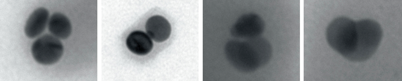
NIST scientists, with collaborators at the University of Michigan, have designed and demonstrated a new and easily tunable, high-contrast scattering agent for biomedical imaging applications. The agent is made from clusters of plates of silver about 50 nanometers across and a few nanometers deep, covered with a polymeric coating – small enough, even when clustered together, to travel into tiny spaces within living cells which are about a thousand times larger.
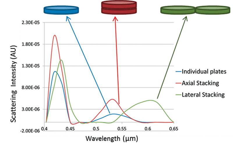
In these light-scattering materials, the nanoplates can exist singly or can cluster into different kinds of stacked orientations inside the polymer coating. In each kind of arrangement, the scientists determined, light scattered from the nanoplates differed sensitively and consistently according to whether the plates occurred singly, aggregated into stacks atop one another (axial clustering), or formed side-by-side arrangements (lateral clustering). Moreover, the measurements agreed well with computer simulations conducted in parallel.
To characterize the nanoplates' behavior, the researchers used a technique called hyperspectral imaging,* in which nanoplate samples were exposed to light at dozens of optical wavelength bands from blue to red (450 nm to 700 nm) at about 10 nm intervals. For each sample, the collected spectra from the scattered light at each wavelength were combined into a "data cube" containing a highly detailed record of optical scattering response across the visible spectrum.
In that manner, the team built up a library showing how the scattering profile was correlated with the stacking orientation. Using the nanoplates in conjunction with the hyperspectral scatter imaging technique, the researchers were able to obtain images of single melanoma cells with 400% better contrast than previously possible. Jeeseong Hwang and Kimberly Briggman of NIST's Physical Measurement Laboratory, along with colleagues at NIST and the University of Michigan, recently reported on the new contrast agent in the journal Biophotonics.**
Why does this work? Noble metals are particularly useful in scattering studies because of the high number of free electrons on their surfaces, which oscillate in response to illumination and determine the scattering profile. Researchers have learned how to bind gold nanoparticles (typically in the form of spheres or cylindrical rods) to specific biological structures and then determine their locations by recording the light that scatters off the particles in response to illumination.
"Eventually, we may be able to use tailored properties of the polymer coating to tag single organelles, individual receptors, mitochondria, parts of the nucleus, and other microstructures," says Hwang, "and then we can determine their location and quantity by measuring the way that each kind of stacking arrangement responds to different colors of light."
If used, for example, to examine samples from endoscopic surgery, they might help determine exactly where the boundary of cancerous tissue lies and thus whether the surgeon has excised all the cancer. Conversely, they could be used to determine the population of pharmaceutical agents.
"When you're developing anti-cancer drugs," Hwang says, "you need to know exactly how much is necessary because too much can kill healthy cells as well. You need to find the optimal concentration. Our nanoplates could, in principle, be used for that."
In the near future, the scientists hope to extend the range of illumination wavelengths into the infrared region beyond about 800 nm. In that case, Hwang says, "we would be able to differentiate very subtle differences and penetrate farther into the cell."
* Whereas an ordinary camera forms an image that combines three broad channels (red, green, and blue), hyperspectral imaging collects emission spectra from dozens or even hundreds of very narrow wavelength bands. NIST has many projects and programs that involve hyperspectral imaging. For more information, click here and here.
** "Scattering based hyperspectral imaging of plasmonic nanoplate clusters towards biomedical applications," A. Ray et al, Journal of Biophotonics, DOI 10.1002/jbio.201500177.