
Official websites use .gov
A .gov website belongs to an official government organization in the United States.
Secure .gov websites use HTTPS
A lock (
) or https:// means you’ve safely connected to the .gov website. Share sensitive information only on official, secure websites.
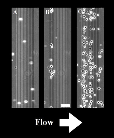
(A) As cells flow down the channel, they pass over the electrodes (vertical dark gray lines) until (B) the electrodes are activated and the cells are trapped and anchored. (C) The cells remain adhered even while being exposed continuously to a fluid flow. Scale bar 50 micrometers.
A research team at the National Institute of Standards and Technology (NIST) has come up with a potential solution to a two-pronged problem in medical research: How to capture cells on a particular spot on a surface using electric fields and keep them alive long enough to run experiments on them.
Their method, which involves innovations upon conventional cell-capture techniques, has already proved effective in creating arrays of human liver cells and mouse pluripotent cells—which, similar to stem cells, can develop into more than one cell type.
"The technique could prove valuable for learning about how cells communicate and differentiate," says NIST chemist Darwin Reyes. "We think this method could provide an effective way to selectively induce cells to differentiate and watch their behavior as they develop."
Adherent cells need to be attached to a surface to survive, and one common way of getting them there is by using a technique called dielectrophoresis (DEP), which Reyes says is not necessarily the best for cells' health. A batch of cells is placed into a fluid medium that has low electrical conductivity—sucrose in water, for example—and then subjected to an electric field that attracts the cells to a nearby surface. But the DEP process requires the cells to spend between 20 and 30 minutes in the medium, which appears to cause problems when the cells are trying to attach to the surface.
"Cells typically die rather soon after that much time exposed to the sucrose, since they cannot attach to the surface," Reyes says. "It's tough to run useful experiments if you only have a short window of opportunity."
The team experimented with different materials before finding that they could use a layer of substance called polyelectrolyte that has its own positive electric charge, which attracts the cells quickly. Before depositing this material, they laid down a thin layer of natural protein called fibronectin that helps cells to survive once they stick. With this new hybrid surface, the cells need spend only about four minutes in the fluid before they are returned to a more nurturing medium that helps them grow and attach better. As a result, the cells can survive on the surface for a week or more.
Because of their success in creating arrays of neural cells, the team has recently started to pattern liver cells as well. Combining liver cells with this technique could be useful in toxicology studies, Reyes suggests. "The liver is made up of several types of cells that work together," he says. "Creating arrays of them with certain cells positioned in particular locations could help us study how each of them might contribute to the overall process of filtering out a toxin from the bloodstream."
D.R. Reyes, J.S. Hong, J.T. Elliott and M. Gaitan. Hybrid cell adhesive material for instant dielectrophoretic cell trapping and long-term cell function assessment. Langmuir, 2011, 27, 10027-10034, DOI: 10.1021/la200762j.