
Official websites use .gov
A .gov website belongs to an official government organization in the United States.
Secure .gov websites use HTTPS
A lock (
) or https:// means you’ve safely connected to the .gov website. Share sensitive information only on official, secure websites.
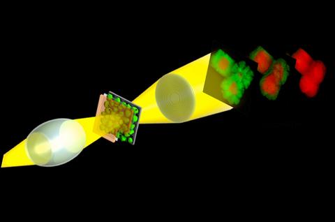
Hard X-ray transmission X-ray microscopy (TXM) is an ideal tool for in situ and operando studies of functional materials and materials synthesis routes. The high energy X-rays provides relatively relaxed restrictions on in situ environments enabling high resolution 2D microscopy and tomography (3D microscopy)1 across a large range of pressures and temperatures and in varying gas or liquid environments. The full field geometry of TXM allows imaging at the sub-second time scale, allowing relevant dynamics to be captured during; for example, battery cycling,2,3 catalysis reactions,4 electrochemical synthesis,5 and corrosion.6 Moreover, by tuning the incident X-ray energy to specific absorption edges, TXM can capture elemental and chemical (spectro-microscopy) changes at 30 nm resolution within a few minutes (Figure).
Li-ion batteries promise the high specific capacity required to replace the internal combustion engine with a number of possible earth abundant electrode materials; however, setbacks such as capacity fading hinder the full capability of these rechargeable batteries. In the search for better electrode materials, high resolution X-ray microscopy during typical battery operation is vital in understand and overcoming the failure mechanisms of these materials. I will discuss the use of X-ray microscopy including spectro-microscopy and nano-tomography to track electrochemical and morphological changes in the electrode material in real time during typical battery operation.
- J. N. Weker, N. Liu, S. Misra, J. C. Andrews, Y. Cui and M. F. Toney, Energy Environ. Sci. 2014, 7, 2771-2777.
- J. Nelson Weker, Y. Li, R. Shanmugam, W. Lai and W. C. Chueh, ChemElectroChem 2015, 2, 1576-1581.
- J. Nelson Weker, A. M. Wise, K. Lim, B. Shyam and M. F. Toney, Electrochim. Acta 2017, 247, 977-982.
- I. D. Gonzalez-Jimenez, K. Cats, T. Davidian, M. Ruitenbeek, F. Meirer, Y. Liu, J. Nelson, J. C. Andrews, P. Pianetta, F. M. F. de Groot and B. M. Weckhuysen, Angew. Chem. Int. Ed. 2012, 51, 11986-11990.
- S. E. R. Tay, A. E. Goode, J. Nelson Weker, A. A. Cruickshank, S. Heutz, A. E. Porter, M. P. Ryan and M. F. Toney, Nanoscale 2016, 8, 1849-1853.
- M. A. Koronfel, A. E. Goode, J. Nelson Weker, S. E. R. Tay, C. A. Stitt, T. A. Simoes, J. F. W. Mosselmans, P. Quinn, R. Brydson, A. Hart, Michael F. Toney, A. E. Porter and M. P. Ryanaccepted to npj Mater. Degrad., 2018, DOI: 10.1038/s41529-018-0029-2.
Speakers
Johanna Nelson Weker (Stanford Synchrotron Lightsource, SLAC National Accelerator Laboratory)