
Official websites use .gov
A .gov website belongs to an official government organization in the United States.
Secure .gov websites use HTTPS
A lock (
) or https:// means you’ve safely connected to the .gov website. Share sensitive information only on official, secure websites.
Addressing Sources of Error in the Cell Viability Measurement Process
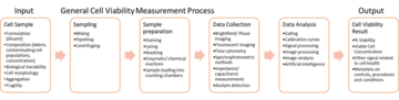
The cell viability measurement process typically involves (1) sampling from a larger volume, (2) sample preparation, (3) data collection, and (4) data analysis (Figure 1). Each step of this process can introduce error into the measurement by either changing the cell samples in ways that can affect the measurement, or biasing results due to the measurement process. We are developing approaches to address different sources of error in the cell viability measurement process.
Measurement Assurance for Trypan Blue-Based Cell Viability Measurements
The trypan blue dye exclusion assay is one of the most commonly used assays for evaluating cell viability. It is based on the principle that viable cells possess intact cell membranes that exclude the trypan blue dye, while non-viable cells have injured membranes and therefore cannot exclude the dye. A cell suspension is mixed with the dye and then immediately visualized under brightfield microscopy (manually or automated) to determine whether cells take up or exclude the dye. A viable cell will appear to have a bright cytoplasm while non-viable cells will appear dark. In recent years, image acquisition and analysis aspects of trypan blue assays have become increasingly automated and studies have demonstrated the improvement in the utility and reliability of these techniques in comparison to manual methods. However, image acquisition and analysis steps in a cell measurement process can introduce several sources of variability that should be controlled to assure measurement quality.
1. Focus Control for Trypan Blue-Based Measurements
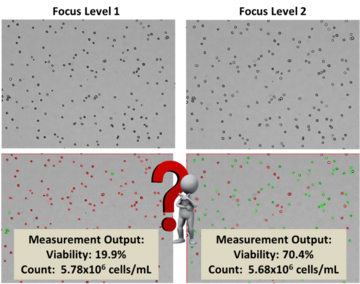
In trypan blue assays, the brightness of objects is highly dependent on the focal plane of imaging. An example of the effect of image focus on a trypan blue dye exclusion cell viability measurement is shown in Figure 2. Analysis of the same field of view of cells collected at two different focal planes with the same image analysis algorithm varied from 19% to 70% cell viability. Upon visual inspection of the images, it is unclear which focus level would be the correct focal plane for analysis, but the difference in the viability result could lead to different decisions being made regarding the usability of the cell sample. This example illustrates the critical need for controlling image acquisition conditions when evaluating cell samples via imaging techniques. Even in cases where image focus is conducted at a fixed focal plane, drifts in instrumentation can cause significant changes in cell appearance, affecting subsequent determination of viable and non-viable cell populations.
Here, we seek to improve the accuracy and precision of trypan blue cell viability assays by implementing control materials to benchmark image quality (e.g. focus and brightness). Introducing a bead material allows for distinct bead features to be analyzed to assess a reproducible focal plane (Figure 3).
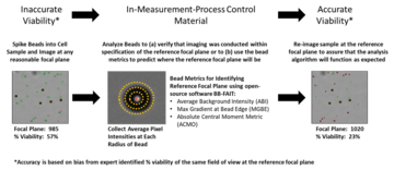
The control beads utilized in these studies were the 100% ViaCheck beads (Bangs Labs, VC50B). This process was demonstrated using the Cellometer Auto 2000 cell viability analyzer. To identify the reference focal plane, using the control beads, an open-source software BB-FAIT (Bead Benchmarking- Focus And Intensity Tool) was developed and trained using a series of training data collected at different focal planes, light intensities, and cell viabilities. Jurkat cells were used as a model cell system. Non-viable Jurkat cells were generated via heat shock treatment (70°C for 30 min) and spiked into nominally viable cells to achieve cell samples at systematically varied % viabilities.
Reference: Peskin, A., Lund, S.P., Pierce, L., Kurbanov, F., Chan, L.L.Y., Halter, M., Elliott, J., Sarkar, S. and Chalfoun, J., 2021. Establishing a reference focal plane using beads for trypan‐blue‐based viability measurements. Journal of Microscopy, 283(3), pp.243-258.
For more information please contact sumona.sarkar [at] nist.gov (sumona[dot]sarkar[at]nist[dot]gov) or laura.pierce [at] nist.gov (laura[dot]pierce[at]nist[dot]gov).
2. Design of Experiments (DOE) for Trypan Blue-Based Measurements
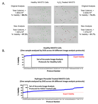
Image analysis is another significant source of measurement error and variability in image-based viability assays. Image analysis algorithms are often automatically included in viability analyzer instrumentation and are sometimes considered proprietary. Parameter values that identify cells and label them as viable or non-viable based on cell features such as size, shape, brightness, sharpness, etc., are routinely used in instrumentation, and these parameter settings can have a significant impact on the reported cell viabilities from the assay (Figure 4A).
We conducted a study in which we systematically varied cell type parameter settings for NIH-3T3 cells analyzed in a stop flow-based trypan blue imaging cell viability analyzer (Vi-Cell XR, Beckman Coulter). Then, we applied a design of experiments (DOE) approach to the image analysis using an orthogonal fractional factorial design to evaluate the sensitivity of the viability measurement to image analysis parameter settings, and to work towards identifying an optimal set of parameters (i.e. image analysis protocols) for evaluating NIH-3T3 cell viability on the trypan blue-based instrument. We found that image analysis parameter settings had a profound effect on the viability reported by the instrument, especially for health-compromised or less viable cell populations (Figure 4B). This study demonstrated both the need to appropriately set image analysis parameters as well as the need to utilize health-compromised cell samples in the optimization of parameter settings.
Reference: Cell & Gene Therapy Insights 2021; 7(4), 551–569 DOI: 10.18609/cgti.2021.076
For more information please contact sumona.sarkar [at] nist.gov (sumona[dot]sarkar[at]nist[dot]gov) or laura.pierce [at] nist.gov (laura[dot]pierce[at]nist[dot]gov).
Contacts
-
(301) 975-8595
-
(301) 975-4370