
Official websites use .gov
A .gov website belongs to an official government organization in the United States.
Secure .gov websites use HTTPS
A lock (
) or https:// means you’ve safely connected to the .gov website. Share sensitive information only on official, secure websites.
Imaging: Neutron Imaging of Lithium and Alkaline Batteries
Summary
A common topic in energy systems is the need for in situ measurement of the mass transport of light ion species such as hydrogen or lithium. Neutron imaging has played a critical role in the understanding of water transport in proton exchange membrane fuel cells (PEMFCs).
Description
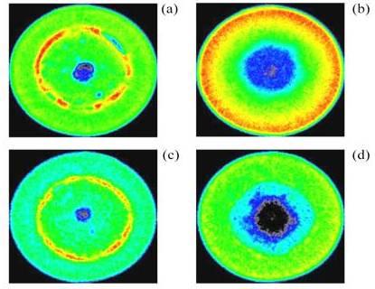
Comparison of the changes in an alkaline AA cell after operation at two different constant currents, were black as low neutron attenuation, red is high neutron attenuation. (a) Fresh cell before operation at 50 mA, (b) discharged cell after operation of 50 mA for 52.5h. (c) fresh cell before operation at a constant 1 A, (d) discharged cell after operation at 1 A for about 70 min. The electrolyte has an initially uniform distribution (green) in the anode (interior) and in the cathode (exterior), and the separator (red/yellow) is clearly visible in (a) and (c).
This is due to the high sensitivity of neutrons to hydrogen, while having relatively small sensitivity to many common materials of construction such as aluminum and carbon. Neutron imaging can play a similar role in advanced battery development by providing in situ measurements of hydrogen or lithium distributions at different states of charge of the battery. The intense, highly collimated, thermal neutron beam has high penetration lengths through battery chemistries of interest, with sufficient sensitivity to measure changes in the ion distribution; by using high resolution imaging detectors, it is possible to resolve features down to 10 μm.
In alkaline primary cells, the neutron attenuation is dominated by the aqueous electrolyte. Tomograms (three-dimensional images) of two AA alkaline cells were acquired before and after discharge to a cell potential of 0 V. Shown in the figure are tomographic slices through two different AA batteries after the end of life achieved by two different current draw conditions, 50 mA and 1 A. In the initial state, the distribution of electrolyte is homogenous in the anode and the cathode. Also, the separator, which is composed of a porous hydrogenous material such as rayon where the pores are filled with electrolyte solution, is clearly visible and is highly attenuating to neutrons. The distribution of the electrolyte after the cell potential has fallen to 0 V clearly depends on the discharge rate. By optimizing particle size to reduce the removal of electrolyte from the anode, it is possible to increase the cell capacity. In more recent experiments, two-dimensional neutron imaging has been used to study a wound prismatic lithium-ion battery. By using high resolution neutron imaging, the thermal expansion of the battery on charge and discharge could be directly measured.