
Official websites use .gov
A .gov website belongs to an official government organization in the United States.
Secure .gov websites use HTTPS
A lock (
) or https:// means you’ve safely connected to the .gov website. Share sensitive information only on official, secure websites.
Summary
In collaboration with Sandia National Laboratories, electric fields have been imaged using neutrons on the NG6e cold neutron imaging beamline. A proof-of-principle experiment to measure the small neutron spin rotation produced by the effective magnetic field produced by the neutron's motion through an electric field was performed. To our knowledge this is the first time an isolated electric field has been nondestructively and directly imaged. This opens the possibility of numerous potential applications, with new capabilities as compared to other electric field methods, in particular the ability to visualize the electrostatic field inside a target volume that can be physically isolated or occupied. To detect the small spin rotation a new scheme for sensitive polarimetry was developed.
Description
An effective magnetic field Beff arises from the relativistic transformation of an electric field E into the frame of reference moving at the neutron velocity v; Beff is proportional to v x E /c2, hence is small due to the factor of the square of the speed of light in the denominator. This effective magnetic field produces a small rotation of the neutron spins in a polarized neutron beam. The overall spin rotation is independent of neutron velocity (and thus also neutron wavelength) because the time spent in the electric field is inversely proportional to the neutron velocity. Hence a full polychromatic neutron beam can be employed, allowing for two orders of magnitude more flux as compared to a typical monochromatic beam. The use of an intense polychromatic beam yields a major advantage for observing the small spin rotation in a reasonable time frame, but a sensitive method for measuring this small spin rotation is still needed. Conventional polarimetry, in which the neutron polarization is aligned with the axis of a polarimeter (like light aligned with an optical polarizer) has low sensitivity to a small tilt of the spin. Hence a key requirement was development of a sensitive but yet simple polarimetry method.
This sensitive method relies on orienting the neutron spin perpendicular to the polarimetry axis. In the proof-of-principle experiment the neutron spin was longitudinal, i.e. along the neutron beam, whereas the polarimeter axis was vertical. This scheme yields the greatest sensitivity to a small tilt of the neutron spin towards the vertical, about two orders of magnitude more sensitive than the conventional method for a typical rotation angle of 0.01 rad (0.6 deg). The polarimetry method developed was facilitated by the established NCNR and PML capabilities in neutron spin filters (NSFs) based on polarized He-3. These devices rely on the highly spin dependent transmission of He-3 gas. The gas is polarized by off-line optical pumping, transported to the beam line, and stored in a vertical solenoid. The key apparatus is a magnetically shielded, vertical solenoid that provides the highly uniform magnetic field required for the He-3 gas while also providing a non-adiabatic transition for the neutron spin. When the neutron passes through the neutron-transparent windings of this solenoid, the laboratory magnetic field changes from longitudinal to vertical very rapidly, thus allowing the neutron spin to be perpendicular to the spins of the He-3 nuclei. A small tilt in this spin induced by the electric field yields a sensitive change in the neutron transmission of the He-3 gas. This solenoid had been developed by the NCNR NSF team for other purposes, but was ideal for the sensitive polarimetry. The method was tested with monochromatic beams on PHADES and NG6e (D.S. Hussey et al, Phys. Procedia 69, 48 (2015)) before being applied to the electric field experiment with the NG6e polychromatic beam.
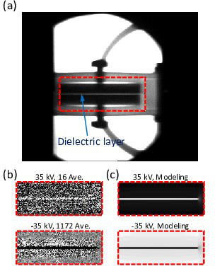
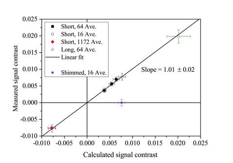
With the polarimetry scheme developed and tested, electric field imaging was accomplished using a high electric field test apparatus developed by Sandia Laboratories. This apparatus provided an electric field of 35 kV across a 5 cm wide, 5.7 cm long, 0.5 mm thick perfluoroalkoxy (PFA) membrane. A neutron image is shown in the Fig. 1(a) and the observed and modelled electric field images are shown in Fig. 1(b) and Fig. 1(c), respectively, for two different numbers of averaged images. As shown in Fig. 2, quantitative agreement was found between the measured and calculated signal contrast for different electric field strengths.
The project was motivated and led by Yuan-Yu Jau of Sandia Labs. The NIST effort was a collaboration between Dan Hussey (instrument scientist for N6e cold neutron imaging beam line), Wangchun Chen (NCNR neutron spin filter program) and Tom Gentile (PML neutron spin filter program). The NG6e imaging beam line infrastructure and the spin filter programs were essential to the success. The electric field results were reported in Y.-Y. Jau et al, Phys. Rev. Lett. 125, 1108 (2020) and the polarimetry method in Y.-Y. Jau et al, Rev. Sci. Instrum. 91, 073303 (2020). This work may enable a new diagnostic technique sensitive to the structure of electric potential, electric polarization, charge distribution, and dielectric constant by imaging spatially dependent electric fields in objects that cannot be accessed by other probes. We expect further collaboration with Sandia on applications of electric field imaging with neutrons.
Notice of Online Archive: This project ended in 2021 and thus this page is no longer being updated and remains online for informational and historical purposes only. The information is accurate as of 5 March 2021. For questions about page contents, please contact Dan Hussey.