
Official websites use .gov
A .gov website belongs to an official government organization in the United States.
Secure .gov websites use HTTPS
A lock (
) or https:// means you’ve safely connected to the .gov website. Share sensitive information only on official, secure websites.
Vector Potential Photoelectron Microscope (VPPEM)
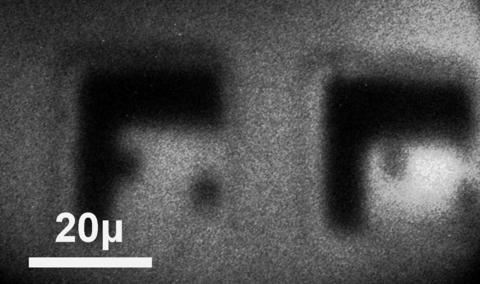
VPPEM image representing the intermetallics in a reaction zone between Aluminum and Calcium is shown in false color.
The Vector Potential Photoemission Microscope endstation sits at the end of both the SST-I and SST-II beamlines, providing both soft (0.1 keV to 2.2 keV) and tender (2.0 keV to 7.5 keV) beams at the sample. The VPPEM is a full field imaging tool based on a new concept in photoelectron microscopy. As with all X-ray Photoelectron Spectroscopy, core electrons with excitation energies below the incident X-ray energy are excited and can be emitted or cause secondary emission in an Auger process. The primary difference in the VPPEM is the method of imaging, where the vector potential, or equivalently the momentum in a magnetic field, acts as a spatial reference instead of a focusing element. Photoelectrons emitted travel down the magnetic field lines until the field is abruptly terminated. Conservation of momentum converts the vector potential into kinetic momentum. Photoelectrons form an angular image based on their point of emission at the sample. The angular photoelectron image is focused into a concentric hemispherical analyzer, energy analyzed, and an image projected onto a detector. Varying the incidental X-ray energy allows the construction of topographic and spectroscopic information such as seen in the figure at the top right.
VPPEM is a technique in development, with known advantages such as the imaging of low energy photoelectrons normally inaccessible to XPS microscopes and the applicability to non-planar samples. As the end station reaches routine operations, further studies hope to evaluate other potential advantages such as the potential for an environmental cell.
VPPEM and the concepts behind the technique were developed through a NIST funded Small Business Innovation Research (SBIR) grant awarded to Ray Browning Consultants. The NIST VPPEM is the only instrument of its type, and is a testbed for the continued development of the technique. Detailed information on the theory can be found on our techniques page (link) and at rbrowning.net.
Specifications/Capabilities
Energy Range: 2.0 keV to 7.5 keV (Tender Beam); 0.1 keV to 2.2 keV (Soft Beam)
Spot Size on Sample: 10 mm to 100 microns
Spatial Resolution: 100 nm to 2 microns (influenced by instrumental magnetic field and surface electric fields of sample)
Depth Resolution at Surface: 1 nm to 10 nm
Sample Environments: Technique applied to surface characterization of solid samples measured in high vacuum. Samples can be non-planar, conducting, or poorly conducting.
Usage Information
Operating Schedule
VPPEM is currently in commissioning and is anticipated to be available to users in 2019.
Access Information
VPPEM is currently in commissioning and is not accepting proposals at this time. After commissioning is complete, access to VPPEM is granted through one of two mechanisms:
1) General users may apply for time through the general user program at NSLS-II. Qualifications, proposal templates, and guidelines are available at https://www.bnl.gov/ps/userguide/.
2) For NIST staff only: NIST projects may request access for experiments by contacting the instrument scientists listed here. Requests are collected and reviewed by the NIST BNL allocation committee and granted time based on merit and fit to the NIST mission. For more information, contact the instrument scientist listed here.