Portable, low-field (1–100 mT) MRI systems that can safely perform scans outside a dedicated MRI suite could revolutionize the use of this diagnostic imaging modality. In addition to alleviating the need for an expensive, MRI-dedicated imaging room, low-field scanners cost far less and require less space and power than traditional MRI scanners that rely on cryogenic superconducting magnets. Such cost advantages make it feasible to deploy low-field MRI scanners in economically challenged hospitals and clinics, while their portability may enable installation in ambulances or portable vans serving remote communities.
The first commercial point-of-care low-field MRI scanner is Hyperfine’s Swoop Portable MR Imaging System, which has CE Mark and US FDA 510k clearance for neuroimaging. Swoop is increasingly used in hospital emergency departments to image patients with severe head trauma or suspected of having a stroke. This portable scanner operates at 64 mT – at least 20 times lower than the magnetic field in conventional MRI scanners.
To expand the clinical application of low-field MRI scanners, however, better contrast agents are needed to improve the image quality. In addition, more research is required to understand the relationship between low-field images and the underlying tissue properties that they represent.
Nanoparticles as contrast agents
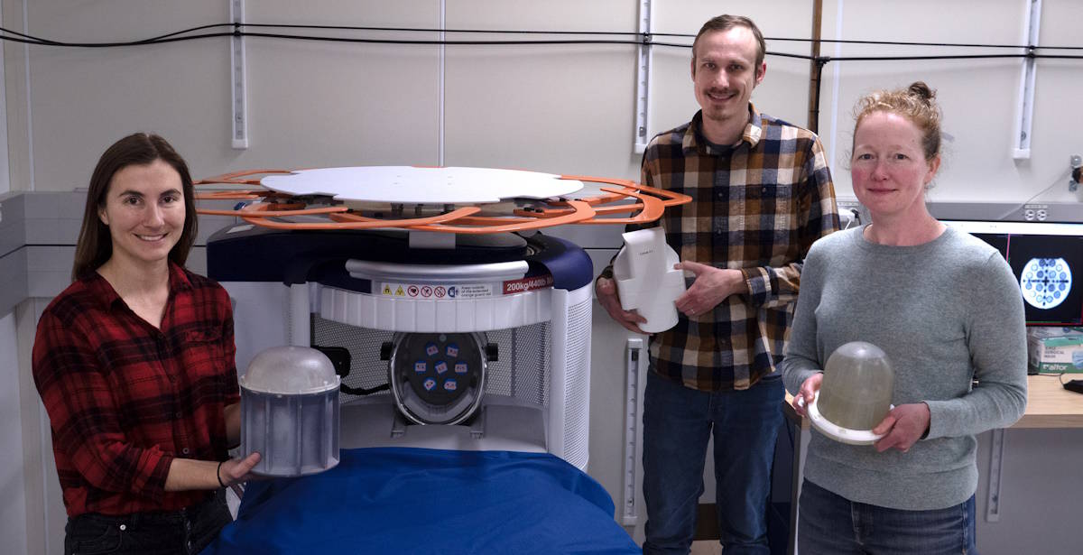
Researchers at the National Institute of Standards and Technology (NIST), the University of Colorado Boulder and the University of Florence have determined that superparamagnetic iron oxide nanoparticles (SPIONs) significantly outperform a commercial gadolinium-based contrast agent (gadobenate dimeglumine, or Gd-BOPTA) used for exams on 3 T MRI scanners. Writing in Scientific Reports, they describe the properties of iron oxide-based contrast agents during acquisition of low-field MRI scans.
Approximately 25% of all MRI exams at clinical field strengths use contrast agents – magnetic materials that are injected into patients to enhance image contrast, enabling anatomical features to be distinguished by their level of brightness or darkness. Contrast agents can help radiologists identify unhealthy tissue based on the MR enhancement patterns of a tumour. For example, the tumour vasculature can accumulate more contrast than healthy tissue, and a tumour that may not have been visible without contrast may become visible.
The efficacy of a contrast agent is directly related to its physical and magnetic properties. Lead author Samuel Oberdick, from NIST and the University of Colorado Boulder, and co-authors characterized monodispersed carboxylic acid-coated SPIONs with diameters ranging from 4.9 to 15.7 nm. Their aim was to understand size-dependent properties of T1 contrast at low field strengths (a T1-weighted MR image demonstrates differences in the longitudinal relaxation times of tissues). By imaging an MRI phantom, they determined the MRI contrast properties at 64 mT using the Swoop system and at 3 T using a preclinical scanner.
The researchers determined that SPION-based contrast agents show favourable qualities as T1 contrast agents for low-field MRI, exhibiting size-dependent longitudinal relaxivities and outperforming Gd-BOPTA by nearly nine times at room temperature and eight times at physiological temperatures. They also observed that the longitudinal relaxivities of SPIONs at 64 mT were nearly an order of magnitude larger than at the standard clinical field strength of 3 T. A high relaxivity enables use of smaller quantities of contrast to create perceptible bright markers on an MR image.

Portable MRI diagnoses stroke at the patient bedside
The team also measured the low-field T1 properties of ferumoxytol, an iron oxide nanoparticle-based treatment for iron deficiency. Ferumoxytol also showed enhanced contrast compared with the gadolinium-based agent. Because it is already FDA approved, ferumoxytol could immediately be used off-label to evaluate the T1 contrast of iron oxide nanoparticle-based contrast agents in clinical studies.
Oberdick advises that the team now plans to explore the optimal properties for SPION-based T1 contrast agents at low fields. Future work may use custom synthesis of nanoparticles to create SPIONs with engineered sizes and magnetic properties to increase T1 contrast at specific low field strengths.
Imaging the brain
Elsewhere at NIST, Kalina Jordanova and colleagues have been working to validate methods for creating images with weaker magnetic fields. They recently measured the properties of brain tissue at low magnetic field strength in a study of five male and five female volunteers, reporting their findings in Magnetic Resonance Materials in Physics, Biology and Medicine.
The team collected 64 mT MR images of the entire brain and obtained data from the grey matter, white matter and cerebrospinal fluid. These three brain constituents respond to the low magnetic field in different ways and produce distinctive signals that reflect their unique properties. This enables the MRI system to produce images containing quantitative information about each constituent.
“With low-field MRI systems, the contrast of the images is different, so we need to know how human tissue looks at these lower field strengths,” says Jordanova. “Knowing the quantitative properties of tissue allows us to develop new image collection strategies for this MRI system,” adds co-author Katy Keenan.