
Official websites use .gov
A .gov website belongs to an official government organization in the United States.
Secure .gov websites use HTTPS
A lock (
) or https:// means you’ve safely connected to the .gov website. Share sensitive information only on official, secure websites.
Summary
Measuring the through-plane water content in a hydrogen fuel cell is critical to understanding this complicated heat and mass transport environment, where the waste heat produces temperature gradients, the inlet and outlet gases are a two-phase flow phenomenon, and the porous media has a wide range of both pore diameters and wettabilities.
Description
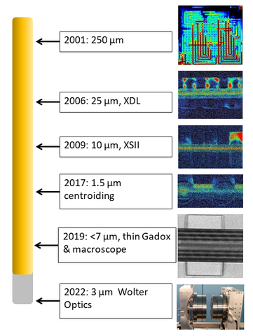
Figure 1: A timeline of neutron detector image spatial resolution realized by the NIST neutron imaging program.
Neutron imaging has yielded valuable insight into the water content of fuel cells because minimal changes to a test section are needed. A limit of neutron imaging is the achievable spatial resolution, which is primarily dominated by the detector. Figure 1 shows the evolution of the spatial resolution of imaging detectors available to NIST and what level of detail can be seen in a hydrogen fuel cell. The Wolter optics system is currently in fabrication. When the neutron imaging of fuel cell project first began, the initial interest was to improve the time resolution in order to capture the dynamics of water flow in the channels of the fuel cell, which was enabled by the use of amorphous silicon flat panel detectors [1].
The approach at NIST is to define critical technical parameters of detectors, collaborate scientifically, and provide support to academic and industrial partners in the development of such detectors. This collaborative effort resulted first in the development of high-resolution detectors based on Micro-Channel Plates (MCPs) with sub-ten micrometer channel diameters. The first generation of readout electronics, based on cross delay line anodes (XDL) was able to reach a spatial resolution of about 25 µm [2]. The second generation of readouts, cross-strip anodes (XSII) [3] is capable of neutron count rates of greater than 4 MHz; this represents a 20-fold improvement in the spatial resolution and factor of 1000 increase in neutron count rate over 4 years and is the current state-of-the-art in neutron detection. While the range of the charged particles in the MCPs limits the current resolution of this technology to about 10 µm, there is still development work in progress to increase the field of view for wider use of the enhanced imaging capability.
Since there is a very significant interest in the studies of water transport, content, and mapping in catalysts layers and membranes, there is also ongoing exploration of other means to further improve the spatial resolution of neutron imaging. To reach spatial resolution less than 10 µm, NIST has developed a macroscope detector to magnify the scintillation light emanating from thin scintillators [4]. With an image intensifier, the macroscope can resolve individual neutron capture events and a centroiding algorithm yields image spatial resolution of better than 1.5 µm [5].
Because the membrane-electrode assembly and many other electrode systems are not readily conducive to simple two-dimensional analysis, improving the time resolution to permit tomography is required. This will be enabled by the neutron microscope based on Wolter optics.
References
[1] M. Estermann and J. Dubois, “Investigation of the properties of amorphous silicon flat-panel detectors suitable for real-time neutron imaging,” IEEE Trans. Nucl. Sci., vol. 52, no. 1, pp. 356–359, Feb. 2005.
[2] D. S.Hussey, D. L. Jacobson, M. Arif, J. P. Owejan, J. J. Gagliardo, and T. A. Trabold, “Neutron images of the through-plane water distribution of an operating PEM fuel cell,” J. Power Sources, vol. 172, no. 1, pp. 225–228, 2007.
[3] O. Siegmund, A. Tremsin, J. Vallerga, and J. McPhate, “Microchannel plate cross-strip detectors with high spatial and temporal resolution,” Nucl. Instruments Methods Phys. Res. Sect. A Accel. Spectrometers, Detect. Assoc. Equip., vol. 610, no. 1, pp. 118–122, 2009.
[4] S. H. Williams et al., “Detection system for microimaging with neutrons,” J. Instrum., vol. 7, no. 2, 2012.
[5] D. S. Hussey, J. M. LaManna, E. Baltic, and D. L. Jacobson, “Neutron imaging detector with 2 μm spatial resolution based on event reconstruction of neutron capture in gadolinium oxysulfide scintillators,” Nucl. Instruments Methods Phys. Res. Sect. A Accel. Spectrometers, Detect. Assoc. Equip., vol. 866, no. April, pp. 9–12, 2017.
Notice of Online Archive: This project ended in 2021 and thus this page is no longer being updated and remains online for informational and historical purposes only. The information is accurate as of 25 March 2021. For questions about page contents, please contact Dan Hussey.