
Official websites use .gov
A .gov website belongs to an official government organization in the United States.
Secure .gov websites use HTTPS
A lock (
) or https:// means you’ve safely connected to the .gov website. Share sensitive information only on official, secure websites.
Performance Benchmarking and Intensity Calibration of a Widefield Fluorescence Microscope
Summary
We have made available a method to characterize a fluorescence microscope's performance by benchmarking the detection threshold, saturation and linear dynamic range to a physical artifact (i.e. a fluorescent material).
Description
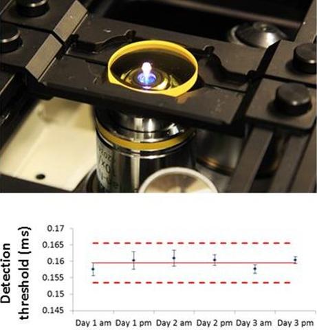
TOP: Image of fluorescent reference material on microscope. BOTTOM: Charting of microscope control parameters.
Widefield fluorescence microscopy is a highly used tool for visually assessing biological samples and for quantifying cell responses. Despite its widespread use in high content analysis and other imaging applications, few published methods exist for evaluating and benchmarking the analytical performance of a microscope. Easy-to use benchmarking methods would facilitate the use of fluorescence imaging as a quantitative analytical tool in research applications, and would aid in instrument validation for commercial product development applications.
To follow this procedure, download and follow this step by step guide.
A script written in MicroManager, an open-source microscopy control software, has been developed to facilitate the implementation of this procedure in other laboratories. The benchmarked parameters can be charted using this Excel Sheet to assure instrument performance over time.
You can watch us implementing this procedure in our laboratory in this short video:
This procedure is available as a Technical Note published in Cytometry Part A.
Acknowledgements: For assistance with the video tutorial, we thank Bryan Goering, Heather Moosher, and Elianna Bier.
*Certain commercial products are identified; this does not imply endorsement or recommendation by NIST.
Major Accomplishments
Beta tested the procedure 4+ institutions.