
Official websites use .gov
A .gov website belongs to an official government organization in the United States.
Secure .gov websites use HTTPS
A lock (
) or https:// means you’ve safely connected to the .gov website. Share sensitive information only on official, secure websites.
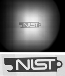
First image taken with NIST's new cold neutron imaging facility. The test sample is a 3 mm-thick stainless steel bottle opener. Top: Raw image. Bottom: Normalized close-up.
Neutrons, the charge-less constituents of atomic nuclei, are nifty imagers. Since the 1950s, scientists have been using these particles' eerie ability to non-destructively penetrate dense matter, creating snapshots – sometimes in 3D – of the insides of fuel cells, rocks, biological samples, and more.
Typical imaging methods involve directing a beam of these particles at a material and then recording how the neutrons interact with atoms in the sample, noting where they are absorbed, scattered away, or pass through unperturbed.
Now a team of researchers at NIST's Physical Measurement Laboratory (PML) and the NIST Center for Neutron Research (NCNR), along with collaborators, have created a new resource for neutron imaging – a "world-class facility, on par with instruments worldwide," says NIST PML's Daniel Hussey. NIST's new amenity is the most recent step in the team's effort to build the world's first practical neutron microscope, which will take high-resolution neutron images in a fraction of the current image-collection time.*
"If the microscope is able to work, it will be a revolution for the field – a factor of a hundred improvement in neutron imaging," Hussey says.
The new imaging facility uses cold neutrons, which have energies of about 5 thousandths of an electron volt (meV, milli-electron volts). Warmer, so-called "thermal" neutrons – the kind used in most nuclear power plant reactors and also for many imaging applications – have energies in the 15-20 meV range and can penetrate more deeply into materials. But cold neutrons generally provide better contrast, have higher sensitivity to water, and are particularly good for measuring the effects of strain within a material.
By harnessing cold neutrons, the new system could help scientists study lithium distribution within batteries, or take before and after photos of the strain in metals. If the material samples are thin enough (on the order of a couple of centimeters), researchers could do high-resolution 3D imaging of the oil and gas in rock samples. The facility might even be used for pharmaceutical research: in a drug processing plant, applying shear to proteins can deform the molecules, which can alter a drug's behavior. Development of new cold neutron imaging methods might provide information about these deformations.
Currently, NIST has demonstrated that they can take cold neutron images with a relatively high resolution, of about 20 millionths of a meter (20 mcm, micrometers).
But to collect this information faster, Hussey explains, the team will need to address one of the major challenges in neutron imaging: achieving a high-intensity, high-resolution beam. And that's where the development of a practical neutron lens could prove useful.
The same abilities that allow these particles to pass unhindered through dense materials like metals – to a neutron, aluminum is basically a window – also makes them difficult to herd. Since they are charge-neutral, researchers can't use magnets or voltage differences to compel them to move anywhere. Instead, neutrons are guided by highly polished and precisely aligned mirrors, which reflect them into the instrument areas, similar to the way a stone might skip off the surface of a lake.
To shine neutrons on a sample, researchers typically direct the beam through a pinhole and illuminate the sample with the beam to create a full image. Any beam that doesn't make it through the pinhole is essentially wasted.
But if those excess neutrons could be redirected onto the sample instead – using a neutron lens based on specially designed mirrors, for example – then the increased intensity of the neutron beams would speed up the image-collection process. That neutron lens is currently being engineered by NIST and collaborators. When it's complete, "what takes us on the order of 20 minutes now will take 20 seconds or less," Hussey says.
Finally, to turn the lens imaging system into a microscope, the team plans to eventually integrate a magnification step into the process, which would increase the spatial resolution by another factor of ten to single-micrometer scale.
While a neutron microscope is a long-term goal to be realized over about three years, PML and NCNR researchers are now focused on improving the density of the neutron beam by two to four times and expanding the system's capabilities to include neutron phase imaging, which uses variations in the intensity of the neutron signal collected by detectors to determine the details of a material's structure at a range of scales, from hundreds of nanometers to about a centimeter.
"We've been losing a lot of sleep to make this all work," Hussey says. "Now that this facility exists, we will begin doing all the cool stuff."
— Reported and written by Jennifer Lauren Lee
*These efforts are part of a 2013 Innovations in Measurement Science (IMS) grant to create a workhorse neutron microscope.