
Official websites use .gov
A .gov website belongs to an official government organization in the United States.
Secure .gov websites use HTTPS
A lock (
) or https:// means you’ve safely connected to the .gov website. Share sensitive information only on official, secure websites.
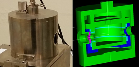
A photograph of the free-air chamber used for the physical measurements (left) and the corresponding model used in the computational simulations (right).
Doctors devising a plan of attack on a tumor may one day gain another tactical advantage thanks to a series of sophisticated calculations proposed by PML's Dosimetry group. Their results could serve as a foundation for the next generation of software used by clinicians, to design courses of radiation treatment for electronic brachytherapy patients.
Electronic brachytherapy is a relatively new cancer treatment similar to conventional brachytherapy, in which radioactive seeds are placed in or near a patient to fight a tumor. In this case, however, the source of radiation is a miniature x-ray tube just a few millimeters in size. Since the tubes use a voltage instead of a radioactive source, they can be turned on and off as needed, allowing clinicians to minimize dose to patients and medical staff.
Currently, doctors use software to decide the best placement for an x-ray tube in a way that maximizes dose to the tumor and minimizes it elsewhere. This software makes several simplified assumptions about radiation dose to the body. For example, traditional brachytherapy dosimetry protocols do not account for the effects of iodine contrast or for the differences in how the radiation interacts with soft tissue and bone. For electronic brachytherapy, these issues can sometimes be significant, resulting in possible discrepancies between the planned dose and the dose that is actually delivered to a patient.
To remedy this situation, PML researchers will perform a series of simulations using state-of-the-art Monte Carlo methods, which rely on a collection of powerful computational algorithms and are often used to model complex physical and mathematical systems. The simulations will incorporate highly detailed generic models of the human body, allowing researchers to study the differences in the absorbed dose received by different organs and tissues.
"Monte Carlo dose calculations are currently considered the de facto gold-standard method for performing patient dosimetry," says PML postdoctoral researcher Matthew Mille, who will be conducting the work. "However, these calculations are much too slow for routine clinical applications." By performing the simulations themselves, the team hopes to provide researchers with the information they need to develop better software tools for physicians, which will allow for more accurate treatment plans for electronic brachytherapy patients.
Before making these calculations, however, Mille will use Monte Carlo methods to assess the performance of the detection system used to make top-level measurements of the radiation emitted by the x-ray tubes.*
For these measurements, the scientists set up an experiment where a tube emits x-rays that are collected by an ionization chamber, in which the interaction of the air and radiation creates a charge in a process known as ionization.
But some of the electrical signal picked up by the detector comes from processes distinct from the radiation emitted by the tube, while other aspects of the signal are lost due to natural processes.** Using Monte Carlo simulations, researchers can calculate how much unwanted ionization contributes to the measured signal.
They use the computed additions and losses as the basis for determining a series of so-called "correction factors," which are then applied to the measurements. These correction factors help doctors ensure that the electronic brachytherapy sources give patients the intended dose.
Unlike previous efforts to determine the correction factors, this latest work will incorporate a more detailed model of the ionization chamber.*** The effort will serve as an independent verification of the older calculations, though the researchers say they anticipate that the new correction factors will be similar to those from earlier results.
-- Reported and written by Jennifer Lauren Lee
*Top-level measurements are performed using primary standard instruments, which are highly accurate and are used to calibrate other instruments.
**For example, some of the x-ray tube's radiation signal goes missing when electrons escape the charge collection region of the free-air chamber before losing all of their energy. On the other hand, some of the electrical signal picked up by the detector comes from processes that shouldn't be included in a measurement of the radiation coming from the x-ray source – for example, bremsstrahlung radiation, created when an electron decelerates.
***The previous set of correction factors was based on Monte Carlo calculations performed at the International Bureau of Weights and Measures (BIPM) and incorporated a simplified model of the ionization chamber as a box instead of a cylinder.