
Official websites use .gov
A .gov website belongs to an official government organization in the United States.
Secure .gov websites use HTTPS
A lock (
) or https:// means you’ve safely connected to the .gov website. Share sensitive information only on official, secure websites.
Medical: Calibrated Phantoms for Monitoring PET and SPECT Scanner Performance in Clinical Trials
Summary
The interpretation of quantitative imaging data obtained from Positron Emission Tomography (PET) and Single-Photon Emission Computed Tomography (SPECT) studies requires an understanding of the measurement variability due to instrumental effects, especially in clinical trials that involve large numbers of patients being scanned in several sites with different types of scanners using a variety of acquisition and analysis techniques. The Radioactivity Group has developed a methodology for traceably calibrating solid epoxy-based phantoms for massic activity and can provide such calibrations on a cost-recovery basis.
Description
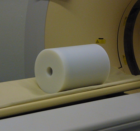
A solid PET calibration phantom filled with Ge-68 in epoxy. The activity concentration is traceable to NIST.
Although the data from most PET and SPECT studies are often used on a relative basis, the quality of the analysis will be greatly improved if the data can be normalized using a common reference standard that allows instrumental variability to be minimized. This requires long-lived phantoms to be available to all sites throughout the entire study and the activity content in the phantoms to be linked to a single standard. When used during scanner commissioning and at regular intervals as part of a complete QA/QC system, these reference phantoms can provide baseline performance metrics for a new scanner and can help monitor long-term performance.
Because the phantoms are calibrated for activity content and are traceable to National standards for radioactivity, they provide “ground truth” for investigating new scanner designs, comparing reconstruction and correction algorithms, and validating analysis software. They can also provide the basis for clinical site validation, accreditation, and performance testing programs.
Working in collaboration with a university medical center and a commercial source manufacturer, we first designed and calibrated two prototype large volume (30 cm length x 20 cm diameter, approximately 9 L) cylindrical phantom sources containing 68Ge in epoxy as a surrogate for 18F that can be used to accomplish these tasks. The design is similar to the phantoms used clinically with 18F to calibrate the PET scanners and are the first ones having a calibrated value for the amount of 68Ge in the phantom that is directly traceable to a national standard for that radionuclide.
Because of their large physical volume, direct calibration measurements on the phantoms were deemed unlikely to satisfy the requirement of having a combined uncertainty on the 68Ge activity concentration of about 1 %. Therefore, a sampling procedure was adopted in which samples of the epoxy containing 68Ge were taken during the phantom preparation process and dispensed into a standardized geometry for calibration. The epoxy sources were measured on several HPGe systems that been calibrated for this specific geometry using a previously standardized 68Ge solution. Corrections were made for possible differences in attenuation and scattering arising from the differences in composition and density between the epoxy and solution sources. From the known mass of the epoxy in each of the samples and these activity measurements, the activity concentration of the epoxy in the phantoms was determined with a combined standard uncertainty of less than 1 %.
Since the initial development of the technique to calibrate the 68Ge phantoms, we have successfully calibrated two large (20 cm to 30 cm diameter) solid 68Ge phantoms for two different manufacturers, thereby providing traceability for their products. We have also applied the technique to smaller source configurations and other radionuclides and have provided calibrations of 68Ge in both the spheres and “warm” background region of a Jaszczak phantom for the UK National Health Service, a set of small (2 mL to 23 mL) cylindrical phantoms for a SPECT image quantification exercise of 133Ba (as a surrogate for 131I) for the International Atomic Energy Agency, and are investigating 75Se as an imaging surrogate for 177Lu.
Many of these phantoms are now becoming commercially available and will provide the nuclear medicine community with the necessary tools to calibrate and monitor the performance of their scanners during clinical trials in a way that is traceable to national standards.