Official websites use .gov
A .gov website belongs to an official government organization in the United States.
Secure .gov websites use HTTPS
A lock (
) or https:// means you’ve safely connected to the .gov website. Share sensitive information only on official, secure websites.
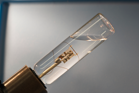
At the center of the cylinder is a microcapillary tube used to study single-particle measurement capability in magnetic contrast agents for MRI.
Thanks to burgeoning progress in magnetic resonance imaging (MRI), it may soon be possible to track and study, in vivo and in real time, heretofore invisible physiological processes – at scales approaching the cellular level – using MRI technology. Eventually, many experts believe, this will include the ability to target and observe multiple specific cell types in action simultaneously.
One critical element in that evolution involves research on tracers called magnetic contrast agents, and PML scientists have recently advanced the field in two critical areas: characterization of conventional contrast agents such as iron oxides, and innovations in an entirely new kind of agent that is microfabricated instead of chemically synthesized.
"There has been enormous improvement in MRI technology over the last 10 years," says John Moreland of the Electromagnetics Division's Magnetics Group, who oversees the contrast work. "And there's much more functional imaging now that is increasingly used in hospitals. As a result, in the industry as a whole, about the same amount of money is now spent on contrast agents as on development and sales of instrumentation.
"There are many contrast modalities, and our group is looking at several. But the common theme is to be able to do real-time imaging with MRI that is analogous to what is now done with positron emission tomography, with much higher resolution and without ionizing radiation."
MRI measures the differences in proton spin relaxation time in water molecules across a range of tissues. A contrast agent has to substantially alter the relaxation properties of nearby protons to the point at which they produce a readily distinguishable signal. Magnetized substances such as gadolinium chelates and various iron oxides have proven useful, but neither is ideal. For example, gadolinium is somewhat toxic, and iron oxide particles provide negative contrast, which is difficult to interpret.
That may be about to change. Moreland and colleagues are pursuing two different strategies designed to reveal different aspects of iron-oxide contrast agents.
One set of experiments involves measuring the way a single magnetic bead – about 1 µm in diameter, made of polystyrene embedded with iron oxide, and placed in a 7 tesla nuclear magnetic resonance (NMR) spectrometer – influences the relaxation time of the tiny volume of water in its immediate vicinity. Those measurements can be used to determine the magnetic moment of the bead. But a major challenge is how to identify the bead-induced signal against the large water background.
To do so, the researchers used a variety of microcapillary tubes, immersed in a surrounding bath of deionized water, with sample volumes (inside the capillary) ranging from 5 nL to 1 nL. Beads were injected one by one, traveling along the tube and through the field of a copper-wire microcoil RF probe wound around the outer circumference of the tube at its center. The scientists then tried different combinations of capillary inner diameter and the number of windings in the coil.
"The goal of this work is to facilitate single-particle measurement capability," Moreland says, "and we've now shown that it's possible to study how single particles behave at the microscale."
The microcapillary results, reported in September of 2012 in the Journal of Magnetic Resonance, showed that the optimal configuration of 1 nL and four windings produced a signal-to-noise ratio of 30, and a volume resolution showing a 600-fold improvement over conventional NMR probes, which have tubes 5 mm long and a 18 µL sample volume.
"No one has ever done a single magnetic-moment measurement in this way," says first author Yoshihiro Nakashima. "By decreasing the sample volume to 1 nL, we can clearly see the difference between the bead effect and the water surrounding the sample."
Another set of experiments uses a novel technique to isolate and measure the magnetic properties of a single particle buried in a composite material using a magnetic force microscope. The instrument's cantilever arm has a 15 µm hard magnetic probe attached to its tip. The arm is suspended over the material containing the 1 µm particle of interest, all of which sits in an external magnetic field of opposite polarity to the probe. The interaction of the two opposing fields results in a small zero-field pocket within the material, and the pocket's location can be precisely controlled by adjusting the distance between the probe and the material.
The researchers sweep the zero-field pocket across the buried particle. By measuring the particle's effect on the cantilever arm's motion, they can determine the response characteristics of the particle as if it were isolated from its surroundings. Moreland and first author Jacob Alldredge published the results in the summer of 2012 in the Journal of Applied Physics, noting that they could determine the buried particle's location to within 0.022 µm.
"The first application of this technology may we be in soil-particle characterization," Alldredge says, "as well as imaging nanomagnetic biomarkers and measuring geologic samples made up of several domains on the micrometer scale."
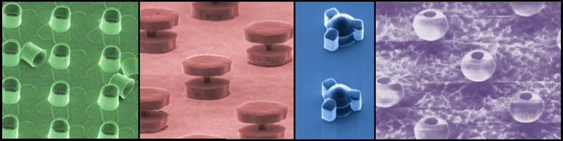
Finally, in a very different and promising approach to contrast agents, Gary Zabow's ongoing experiments on microfabricated contrast agents have produced an intriguing new variation on landmark work that he and colleagues at NIST and the National Institutes of Health first reported in a 2008 paper in Nature. That research showed that micro-engineered magnetic structures – in contrast to chemically synthesized particles – produce readily distinguishable RF spectra, and that the specific spectral characteristics are determined by each structure's geometry. Those differences can be represented in MRI imaging as different colors.
"One possible application is cell targeting," Zabow says, "which is one of the things we want to do – track cells as they move throughout the body, or track different biomarkers."
Zabow generally has worked on two basic structures, an open cylinder and a pair of parallel magnetic disks separated by a non-magnetic connector. "But recently we've demonstrated a new range of structures. They look a little weird, but potentially can be made much cheaper without a lot of the expensive clean-room equipment that we currently use. That ellipsoid shape, which is something like an eggshell with one end removed, doesn't necessarily require the kind of costly photolithographic patterning used in our other shapes."
Whatever the shape, Magnetics Group leader Ron Goldfarb believes the technology has enormous promise: "Let's say you were doing a screen for various disease possibilities. You could have a cocktail of different micro-engineered nanoparticles, each one functionalized differently with a distinctive coating that enables it to target a different type of cell. When each type of nanoparticle gets to its target, it exposes the surrounding water to a field that's different from the fields of the other nanoparticles at their specific targets. You would be able to image all of this at once in MRI, and what you would see is very different shifts in the proton resonance frequency for each particle type. You could assign 'colors' to each, and end up with a comprehensive image."