
Official websites use .gov
A .gov website belongs to an official government organization in the United States.
Secure .gov websites use HTTPS
A lock (
) or https:// means you’ve safely connected to the .gov website. Share sensitive information only on official, secure websites.
Summary
The sizing and counting of subvisible particles in the size range 2 μm to 100 μm is an essential component of product quality assurance in therapeutic protein formulations. Particles may originate from aggregation of the protein, or from non-proteinaceous sources such as the packaging, silicone oil, and other contaminants. Of particular concern are particles from protein aggregates as their presence can contribute to an immunogenic response as well as affect the potency of a drug. Characterization tools for particles in this size range include light obscuration (LO), flow microscopy (FM), and electrical sensing zone (ESZ). While generally these methods agree with one another when measuring standard reference spherical beads, reports on measurements of particles in protein formulations often provide significantly different results. To resolve these discrepancies, this project uses a microfluidic approach to characterize particles simultaneously using two or more techniques, for example FM and ESZ. Lithographically designed particles will be used as models to clarify particular aspects of the measurements.
Description
Intended Impact
The results of this work should provide instrument manufacturers with data, methods, and algorithms for enhancing the clarity and accuracy of information their instruments provide to their customers. Users of these instruments (pharma companies) and regulators, will have better understanding of the factors to consider when comparing results on different types of instruments. In addition, the research may lead to new microfluidic instruments that provide more comprehensive data using smaller sample sizes.
Objective
The primary objective is to identify the factors that influence the parameters reported by different measurement techniques. Simultaneous measurement of particle morphology, electrical signal, fluorescence, etc. on each particle produces a data set that will facilitate comparison of results using these techniques separately. In addition, the research will test the capabilities for microfluidics to perform comprehensive subvisible particle analysis on small samples.
Technical Approach
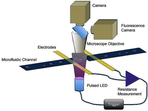
A microfluidic device consisting of a microchannel with four electrodes is mounted in a fluorescence/optical microscope. The microscope is outfitted with fast, computer-controlled cameras (both optical and fluorescence) and with a bright 480 nm light emitting diode(LED). The microchannel features an electrical sensing zone, between two electrodes, where the channel width is reduced.
| When a particle enters this zone, the electrical signal triggers the exposure of the cameras. A controlled delay with microsecond resolution is used to trigger the flash of the LED which lasts typically 5 ms. The camera produces an image which is correlated with the electrical signal. Both camera images and electrical signals are stored for each particles analyzed. Fluorescence imaging also makes use of optical filters to isolate fluorescence from labeled particles. By use of multiple flashes during a single exposure, a strobe-like image is obtained of the particle's trajectory through the microchannel. Image-processing software analyzes the particle images for size, aspect ratio, brightness, etc, while peak analysis software analyzes the electrical signals. |
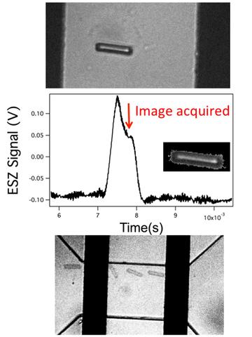 Top: 30 mm long rod in microchannel, middle ESZ signal correlated with image time, inset software-isolated particle used in calculating dimensions, bottom: strobe image of rod tumbling in a microchannel |
Major Accomplishments
- Simultaneous acquisition of image and electrical signal of standard reference beads
- 30 particles per second maximum acquisition rate achieved
- Strobe imaging provides correlates particle trajectory with signal for beads
- Lithographically manufactured particles show shape effects on signal size, and tumbling revealed in stroboscopic measurements
- Signals and images from protein particles obtained
- Fluorescent beads captured with simultaneous to optical and electrical signal
Presented Work
Michael Carrier -IFPAC 2013 Session on Particle Characterization Engineered Particles for the Characterization of the Performance of Protein Particle Analysis Instruments, Jan. 25.
WCBP: 17th Symposium on the Interface of Regulatory and Analytical Sciences for Biotechnology Health Products, Jan. 29-31. "Comparison of particle analysis tools using engineered particles as simulants for protein aggregates"
Invited talk at 5th Annual The Bioprocessing Summit "Comparison of Methods for Characterizing Subvisible Particles Using Manufactured Particles and Microfluidics" Boston, MA, Aug. 21.
Michael Carrier-Invited IBC's 17th Annual Well Characterized Biologicals, October 21-22, "Understanding the response of particle detection instruments to non-spherical particles" 2013